
Quick Summary: Cherry angiomas are common, completely benign vascular skin growths consisting of small, dense collections of blood vessels. They pose absolutely zero risk to your physical health, requiring no medical treatment beyond optional tissue-sparing clinical procedures for cosmetic preference or symptom control.
What Is Causing These Small Red Bumps?
Understanding the natural genetic signaling pathways of your blood vessels is a strategic first step in managing cherry angiomas. These growths occur when the small cells lining the inside of your blood vessels (endothelial cells) multiply rapidly in a tiny, highly localized cluster. You can think of a cherry angioma as a completely safe, localized “vascular landmark” rather than a dangerous or erratic cellular mutation.
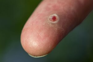
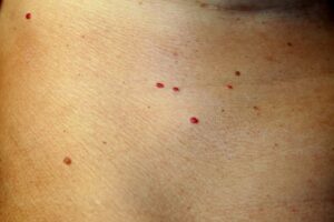
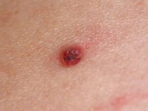
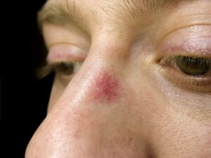
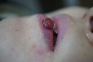
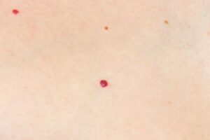
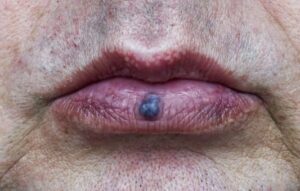
The root cause of these stable papules is a spontaneous genetic shift in somatic cells, specifically within the GNAQ or GNA11 genes. Identifying these bright landmarks early is the “So What?” factor in your skincare routine, giving you complete reassurance that they are non-infectious, structurally stable, and entirely unrelated to internal vascular disease.
Understanding the Presentation: Standard vs. Thrombosed Angiomas
Cherry angiomas can shift in their visual profile due to minor mechanical factors. Distinguishing between these two states prevents unnecessary worry during self-skin checks.
| Vascular State | Key Characteristics and Appearance |
|---|---|
| Standard Cherry Angioma | Presents as a small, firm, dome-shaped bump with a uniform bright red, deep red, or purple hue. Range in size from 0.1 to 1 cm. |
| Thrombosed Angioma | Occurs when a tiny blood clot forms inside the growth, often following minor scratching. This causes the bump to turn dark blue or black, mimicking dark moles. |
Am I at risk for cherry angiomas?
Developing cherry angiomas is an incredibly widespread aspect of the body’s natural maturation process. They are entirely independent of skin cleanliness, dietary choices, or environmental toxins.
- The Age Threshold: Susceptibility expands significantly with time. The total number and overall size of these bumps increase dynamically after the age of 40, affecting an estimated 75% of individuals over the age of 75.
- Hormonal Influences: It is common for cherry angiomas to erupt suddenly or expand in number during pregnancy due to systemic vascular growth signaling.
- Genetic Traits: Family history plays a strong role; having a parent with extensive angiomas often predisposes you to a similar skin pattern.
Where and How It Appears on My Body
Cherry angiomas follow a highly scattered, non-patterned “map” across the body surface that helps your provider instantly recognize them during a full-body skin exam.
- Trunk Dominance: They show a profound preference for the chest, back, abdomen, and shoulders, presenting as multiple separate points.
- Anatomic Sparing: It is exceptionally rare for classic cherry angiomas to develop on the mucous membranes or the skin of the hands and feet.
- The Crimson Profile: Look for a distinct crimson or ruby coloration. On dark skin tones, they can reflect light beautifully as firm, deep purple or ink-like nodes embedded smoothly in the skin.
Solutions I Can Try at Home
Because cherry angiomas are vascular structures fed directly by underlying capillaries, the safest at-home strategy is a gentle, hands-off routine.
- Do Not Pick or Scratch: Trying to squeeze, pick, or scratch off an angioma will not eliminate it. Because it is a dense collection of blood vessels, scratching will result in sharp, sudden bleeding and can introduce local bacteria.
- Grooming Protection: If you have raised angiomas in areas where you shave (such as the face, neck, or limbs), take extra care with your razor. Shaving nicks on an angioma can cause persistent bleeding that requires a clean bandage and firm, continuous pressure for 10 minutes to stop.
- Observe and Reassure: Keep your skin hydrated and celebrate its stability. Rest assured that these safe growths remain stable without transforming into systemic complications.
When Should I See a Dermatology Provider?
While treatment is completely optional, seeking professional validation is recommended if a lesion changes its characteristic features or causes anxiety.
Seek Professional Help if You Notice These “Red Flags”:
- Diagnostic Uncertainty with Black Bumps: An angioma turns black or dark purple (thrombosed). While this is usually a benign clot, a dermatologist should examine it with a dermatoscope or perform an in-office punch biopsy to rule out a pigmented basal cell carcinoma or a melanoma.
- Rapid Modification or Pain: An isolated red nodule grows unusually large, becomes tender, or bleeds spontaneously without a known scratch, which requires evaluation to rule out a pyogenic granuloma or an amelanotic melanoma.
- Cosmetic Clearance: The spots cause aesthetic distress, and you wish to explore precise, scar-free in-office removal methods like electrosurgery or a specialized vascular laser.
Frequently Asked Questions
- Q: What happens if a provider examines an angioma with a dermatoscope?
A: Under a dermatoscope (a specialized magnifying skin instrument), a benign cherry angioma reveals a highly specific, comforting visual hallmark known as a “red-clod” or “lacunar” pattern. This structures looks like a cluster of tiny, red, rounded spaces, immediately setting it apart from dangerous pigment networks. - Q: Will removing an angioma cause more to erupt around it?
A: No. Removing an angioma has zero effect on the adjacent skin cells and will not stimulate new growths. However, because your internal genetic blueprint controls vascular development, your skin may naturally continue to form separate angiomas elsewhere as you grow older. - Q: Why does my provider discourage home freezing kits for angiomas?
A: Over-the-counter wart kits are designed for superficial warty skin tissue. Using them on a vascular lesion like an angioma carries a high risk of causing an accidental chemical burn, persistent bleeding, or permanent color loss (hypopigmentation) that is far more prominent than the original small red spot.
The long-term outlook for angiomas is outstanding, as they are entirely benign skin variations that present zero risk to your health. Success lies in annual professional validation, gentle skin habits, and choosing precise, tissue-sparing clinical removal methods if you desire cosmetic refinement.