Basal Cell Carcinoma (BCC) is the most common form of skin cancer and the most frequently diagnosed of all cancers worldwide. Arising from the basal cells at the bottom of the outer skin layer, BCC is primarily caused by long-term, cumulative exposure to sunlight. While it rarely metastasizes to vital organs, it can cause extensive damage to surrounding tissues if left untreated. Early diagnosis via a skin biopsy and prompt surgical management are critical milestones to eliminate the malignancy, optimize cosmetic outcomes, and safeguard your health.
What Is Causing This Type of Skin Cancer?
Understanding how environmental triggers interact with your skin’s cellular lifecycle is a strategic first step in managing your diagnosis. Basal cell carcinoma originates in the basal cells, which are located at the bottom of your outermost skin layer, the epidermis. This condition is entirely independent of personal cleanliness, hygiene flaws, or surface allergies.
The primary driver of almost all basal cell carcinomas is chronic, cumulative exposure to ultraviolet (UV) radiation from sunlight. Over years of exposure, UV light damages the DNA within your basal cells, disrupting their normal lifecycle and pre-programming them to multiply uncontrollably rather than shedding normally.


Podcast:
Learn More About Basal Cell Carcinoma
While sun exposure is the overwhelming primary cause, rare contributing factors can trigger the disease process in unexposed areas. These secondary engines include chronic contact with arsenic, exposure to ionizing radiation, or long-standing complications arising at the site of old burns, scars, vaccinations, or even tattoos. You can think of a BCC as a localized “cellular coding error” prompted by cumulative radiation stress. Identifying these growths early through an expert clinical evaluation is the essential “So What?” factor in your skincare routine. Catching a BCC when it is small simplifies treatment, prevents deep tissue penetration, and ensures an excellent long-term cosmetic cure.
Understanding Your Skin Status: Comparing BCC to Common Pre-Cancers
Because basal cell carcinoma can present as a scaly or reddish spot on sun-damaged skin, it is frequently confused with pre-cancerous lesions like Actinic Keratosis (AK). Recognizing the distinct clinical and structural differences between these two conditions avoids medical delays and ensures you receive the correct level of care.
| Skin Lesion Type | Key Differentiators and Structural Surface Layouts | |
|---|---|---|
| Actinic Keratosis (AK) |
| |
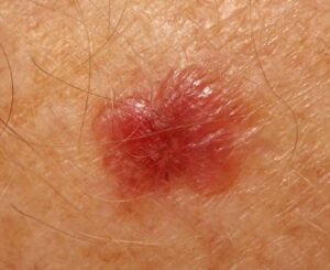
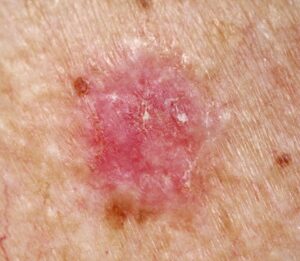
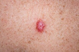
| Basal Cell Carcinoma (BCC) |
|
Am I at risk for Basal Cell Carcinoma or future recurrences?
Developing basal cell carcinoma is directly tied to lifetime ultraviolet radiation exposure and specific genetic traits. Anyone with a personal history of sun exposure can contract this condition, but distinct profiles alter your baseline risks.
- The High-Risk Phenotype: Individuals possessing fair skin, blonde or red hair, and blue, green, or gray eyes carry an elevated genetic susceptibility to solar damage.
- Occupational and Geographic Multipliers: Workers in occupations requiring long hours outdoors and people who spend extensive leisure time in the sun are particularly susceptible. Geographic location is a massive statistical factor: the closer you live to the equator, the higher the total number of documented cases due to intense UV rays.
- The Changing Age Axis: While BCC remains rare in children, it occasionally surfaces in teenagers. Furthermore, skin specialists report a concerning trend where more and more young adults in their twenties and thirties are being actively treated for skin cancer.
- The Two-Year Recurrence Trajectory: Having one documented BCC places you at an automatic lifelong risk for developing others, either at the original site or elsewhere on your body. Statistically, the vast majority of recurrences take place within the first two years following surgery, making structured surveillance critical.
Where and How It Appears on My Body
BCC landmarks map out across highly predictable anatomical corridors, seeking out the specific zones that receive the maximum volume of direct sunlight.
- The Sun-Exposed Anchor: Tumors occur most frequently on chronically exposed landscapes, populating the face, ears, neck, scalp, shoulders, and back.
- The Scalp and Nose Risks: While BCC can develop anywhere, lesions arising directly on the scalp and the nose are historically documented to be **especially troublesome** and carry a higher risk of local complexity or recurrence.
- The Five Visual Warning Signs: Tracing a basal cell growth typically reveals one or more of these classic architectural footprints:
- An Open Sore: An unyielding sore that bleeds, oozes, or crusts, remaining open for weeks only to heal superficially and bleed again.
- A Reddish Patch: A flat, irritated red area occurring on the chest, shoulders, arms, or legs that may itch or crust.
- A Pink Growth: A smooth, slightly elevated pink bump with a slightly crusted central indentation and tiny visible blood vessels.
- A Shiny Bump: A translucent, pearly nodule that can appear pink, red, or clear, often confused with a common mole on deeper complexions.
- A Scar-Like Area: A firm white, yellow, or waxy area with poorly defined borders, resembling a scar, which often signals an aggressive, infiltrating tumor architecture.
- Muted Complexion Clues: On deeper skin tones, the classic pearly translucent bump may lack a bright pink background, appearing instead as a shiny brown, black, or darkly pigmented nodule that can mask itself as a benign mole or seborrheic keratosis. On skin of color, checking for a firm, waxy texture and looking closely for fine, thread-like telangiectatic vessels under good lighting remain vital hooks to identify active changes.
Solutions I Can Try at Home
Because basal cell carcinoma is an active, established skin malignancy driven by internal DNA damage, home remedies, topical creams, or natural poultices provide zero structural benefit and can cause dangerous medical delays. At-home support focuses entirely on mechanical screening and preventing further light-driven cellular mutations.
- Perform Meticulous Monthly Self-Exams: Examine your skin regularly—at least once a month—if you are in a high-risk category. Utilizing a full-length mirror and a hand-held mirror in a well-lit room, systematically inspect hard-to-see areas including your scalp, the backs of your ears, your lateral neck, and your back.
- Implement Strict Daily UV Field Protection: Apply a broad-spectrum sunscreen daily, wear wide-brimmed hats, and protect your skin with tightly woven sun-clothing. Preventing new radiation breaks supports your global skin health and shields adjacent sun-damaged tissues from developing secondary malignancies.
- Never Attempt to Scratch or Pick the Growths: If you spot a scabby bump or an open sore, do not pick at the crust or try to scratch the lesion off. Because the cancer cells reside deep within the basal layer, manual irritation will only cause localized bleeding and painful inflammation without removing the tumor.
When Should I See a Dermatology Provider?
BCC cannot be definitively diagnosed by sight alone and requires expert clinicopathological correlation. Seeking professional clinical triage early ensures you secure an accurate diagnosis via a safe, in-office diagnostic skin biopsy to evaluate your tissue layers under a microscope and rule out aggressive mimics.
Consult Our Office Immediately if You Notice These Warning Signs:
- The Non-Healing Open Sore: You observe a spot, sore, or crusted patch that bleeds unprompted, fails to heal completely within 3 to 4 weeks, or repeatedly heals and breaks open again.
- A New Pearly, Shiny, or Changing Bump: A translucent, pink, smooth nodule emerges on your face, nose, ears, or scalp, or an old skin spot begins to shift in size, color, or thickness.
- The Appearance of an Unexplained Scar-Like Area: You notice a firm, taut, white or yellow scar-like patch developing in an anatomical region where you have never suffered a physical tissue injury or prior surgery.
Frequently Asked Questions
- Q: What primary in-office surgical options are used to eradicate standard basal cell carcinomas?
- Fortunately, there are several highly effective surgical modalities available to cleanly eradicate BCC. For standard lesions on low-risk areas, your provider can execute Excisional Surgery, where they remove the entire growth along with an additional border of normal skin called a “safety margin,” closing the site with stitches and sending the tissue to the lab to verify clean margins. Alternatively, for small or superficial tumors, your provider can perform Curettage and Electrodesiccation, scraping the cancerous tissue away with a sharp, ring-shaped instrument called a curette and utilizing an electric needle to cauterize residual cells and control bleeding. These procedures are performed comfortably under a localized numbing anesthetic on an outpatient basis.
- Q: What is Mohs Micrographic Surgery, and when is it recommended for my care?
A: For tumors located on high-risk cosmetic or functional areas—such as the face, nose, eyelids, lips, or ears—or for large and recurrent growths, Mohs Micrographic Surgery is the gold-standard treatment. During this precise technique, your provider removes very thin layers of the malignancy, layer by layer, mapping and checking each one under a microscope right in the office. The excision is repeated strictly until the tissue slices are verified to be completely tumor-free. Mohs surgery saves the maximum amount of healthy skin and delivers the highest statistical cure rate of any treatment modality. - Q: What is the recommended long-term clinical checkup schedule following my diagnosis?
A: Because having one basal cell carcinoma places you at an elevated risk for developing additional skin cancers, regular clinical health maintenance is mandatory. The Skin Cancer Foundation advises having a thorough, total-body skin examination by a qualified specialist at regular intervals. Depending on your specific skin type, history of sun exposure, and the number of active lesions, your physician will suggest a customized time frame for follow-up visits—typically scheduling checks **every 3 to 6 months** initially if you have widespread sun damage, with the ultimate goal of maintaining meticulous **annual total-body examinations** once control is achieved.
The long-term outlook for individuals managing basal cell carcinoma is outstanding when tumors are identified and treated promptly in their early stages. Success relies on maintaining strict monthly self-examination habits, practicing daily broad-spectrum sun field protection, and adhering to your scheduled follow-up checkups with your interprofessional dermatology team to keep your body safe, healthy, and fully protected.