
Actinic Keratosis (AK), also known as solar keratosis, is a common precancerous skin condition characterized by rough, scaly spots that develop on areas of the skin heavily damaged by long-term sun exposure [cite: 1391, 1479]. While an individual spot can look like a simple patch of dry skin, AKs represent the earliest clinical step in the development of squamous cell carcinoma (SCC), a common form of skin cancer [cite: 1438, 1480]. Understanding the layout of these spots through sensory exams, protecting your skin from further damage, and utilizing field-directed or targeted removal methods are crucial milestones to clear active lesions, repair your cellular barrier, and protect your long-term health [cite: 1415, 1448, 1475].
Podcast:
Learn More About Actinic Keratosis
What Is Causing These Rough, Scaly Spots?
Understanding how cellular changes and environmental stress interact within your skin layer is a strategic first step in managing actinic keratosis. The primary cause of AKs is direct DNA damage to your skin cells (keratinocytes) provoked by short-wavelength ultraviolet B (UVB) radiation from natural sunlight or artificial tanning beds [cite: 1400]. When your skin absorbs this cumulative radiation over many years, it causes mutations that disrupt the pre-programmed growth cycle, leading to abnormal, rapid skin cell development [cite: 1400].
This localized process is often accelerated if an individual has poor immune function due to advanced age, recent heavy sun exposure, systemic disease, or specific medications [cite: 1401]. You can think of an actinic keratosis as a tiny “cellular factory error”—where damaged skin cells multiply improperly and accumulate on the surface rather than shedding normally [cite: 1400]. Identifying these rough textures early through professional clinical exams is the absolute “So What?” factor in your skincare routine [cite: 1414, 1415]. Because approximately 10% of active, red, and tender lesions will take the next step and progress to an invasive squamous cell carcinoma, early intervention safely eliminates the precancer before it can penetrate deeper tissue or metastasize [cite: 1439, 1474].
Understanding Your Skin Status: Comparing AK to Widespread DSAP
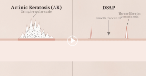
Because actinic keratosis creates rough, red patches on sun-damaged limbs, it is frequently confused with another chronic sun-induced condition called Disseminated Superficial Actinic Porokeratosis (DSAP) [cite: 1433]. Recognizing the key physical differences prevents clinical confusion and directs your provider toward the right treatment path [cite: 1414, 1434].
| Skin Condition | Key Differentiators and Structural Behaviors |
|---|---|
| Actinic Keratosis (AK) | Presents as individual or grouped red, pink, or brownish plaques with an intensely rough, gritty, sandpaper-like texture [cite: 1392, 1393]. They lack a distinct raised border line and are characterized strictly by a prominent, adherent rough scale sitting on an inflamed base [cite: 1392, 1510]. |
| Disseminated Superficial Actinic Porokeratosis (DSAP) | Presents symmetrically on the lower limbs and forearms as flatter, significantly smoother patches [cite: 1433, 1434]. Instead of a gritty surface scale, each spot is bounded by a highly unique, slightly elevated thread-like ridge or rim known as a cornoid lamella [cite: 1384, 1434]. |
Am I at risk for actinic keratosis or skin cancer progression?
Developing actinic keratoses is an environmental event driven by cumulative radiation exposure. It has absolutely zero connection to personal hygiene flaws or poor skin cleanliness [cite: 1400].
- The Lifelong Exposure Profile: Approximately 1 in 6 individuals will develop AKs over their lifetime, with the risk multiplying if you live in tropical or subtropical climates, have a history of frequent blistering sunburns, or spend long hours working or playing outdoors [cite: 1403, 1405, 1408, 1409].
- The Fair Skin Phenotype: Individuals possessing fair skin, blue, green, or gray eyes, blonde or red hair, and a tendency to burn easily before tanning possess an elevated genetic susceptibility to UVB-induced DNA damage [cite: 1407, 1411, 1412, 1413].
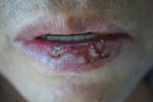
- The High-Metastatic Lip Threat: When actinic damage populates the vermilion border of the lower lip, it manifests as diffuse scaling, dryness, or erosions called **actinic cheilitis** [cite: 1398, 1440]. This is the most aggressive form of keratosis; if it progresses to squamous cell carcinoma, it carries an alarmingly high metastatic rate of 16% to 20% [cite: 1427, 1441].
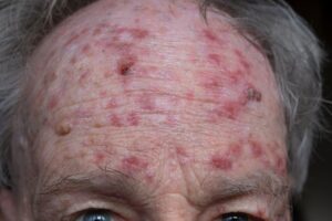
- The Field Cancerization Risk: Having widespread plaques across your face or scalp signifies “field cancerization,” where broad areas of normal-appearing skin harbor hidden, sub-clinical precancerous cell changes that require extensive treatment [cite: 1448, 1449].
Where and How It Appears on My Body
AKs populate highly predictable maps across your body, seeking out the specific landmarks that receive the maximum volume of direct sunlight [cite: 1396].
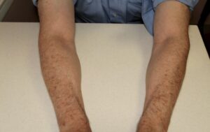
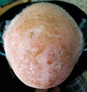
- The Sun-Exposed Corridor: Spots develop heavily across the backs of the hands (dorsal hands), forearms, the face, ears, the lateral neck, and the thinned or balding scalp [cite: 1396, 1491]. In patients with extensive, severe photoaging, lesions will expand onto the upper trunk, lower legs, and the tops of the feet [cite: 1397, 1406].
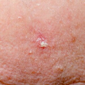
- The “Feel Better Than See” Rule: A cornerstone of identification is that an AK can often be felt before it can be seen clearly [cite: 1415]. Running a finger gently across sun-damaged skin reveals a sharp, coarse, rough texture that contrasts sharply against the surrounding smooth tissue [cite: 1415, 1416].
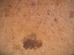
- The Multi-Color Scaling Profile: Lesions vary from 1mm to 2cm in size and present as skin-colored, pink, bright red, or dark brown papules [cite: 1393]. They feature a white, yellow, or grimy crusted scale that can occasionally grow into a hard, horn-like projection known as a **cutaneous horn** [cite: 1392, 1424, 1482].
- Muted Complexion Clues: On deeper skin complexions, the background redness may look muted, hyperpigmented dark brown, or gray [cite: 1393]. In skin of color, the rough, gritty sandpaper texture remains the primary clinical anchor used to locate active spots [cite: 1392, 1415].
Solutions I Can Try at Home
Because actinic keratoses are active precancerous growths driven by internal DNA damage, home care focuses heavily on aggressive daily UV protection to cut off the radiation fuel source [cite: 1400, 1476].
- Implement Strict Sun Protection: This is your single most vital preventative tool [cite: 1476]. Apply broad-spectrum sunscreen daily, wear wide-brimmed hats, and protect your arms with tightly woven clothing [cite: 1476]. Strict sun avoidance prevents new DNA breaks and helps your body’s immune surveillance pathways manage early cell anomalies [cite: 1401, 1476].
- Avoid Aggressive Exfoliation: Do not use rough loofahs, harsh pumice stones, or strong body scrubs to force off the thick, gritty scales [cite: 1392, 1450]. Because the scale is generated from a clone of atypical epidermal cells rather than surface dirt, scrubbing breaks the skin barrier, causes painful inflammation, and does not stop the underlying precancer [cite: 1400, 1453].
- Nicotinamide and Topical Triage: Discuss adding oral nicotinamide (Vitamin B3) with your clinical team to help support cellular energy lines and skin repair [cite: 1467]. However, note that nicotinamide should be avoided if you have a prominent history of major cardiovascular events to ensure your complete physical safety [cite: 1467].
When Should I See a Dermatology Provider?
Actinic keratosis cannot be safely diagnosed by sight alone if it begins changing. Seeking professional clinical triage is necessary to determine if a lesion has crossed the boundary into a malignant skin cancer, which requires an expert **shave or punch skin biopsy** extending down into the superficial dermis to evaluate the core architecture [cite: 1417, 1418].
Seek Professional Help immediately if You Notice These Warning Signs:
- The Rapidly Changing Plaque: A suspected sun spot rapidly expands, becomes significantly thicker, turns exquisitely tender or painful, or breaks open into an open, non-healing sore or ulceration, indicating a progression toward squamous cell carcinoma [cite: 1426, 1430].
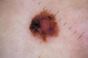
- The Large or Horn-Like growth: You develop an atypical lesion larger than 7mm, notice a firm warty projection (cutaneous horn), or have diffuse scaling and cracking on your lower lip (actinic cheilitis) [cite: 1423, 1424, 1427].
- The Treatment Failure Node: A spot has been previously treated with liquid nitrogen or prescription creams but completely fails to clear up or recurs rapidly at your follow-up checkup, requiring an urgent biopsy to evaluate the deep tissue base [cite: 1417, 1421].
Frequently Asked Questions
- Q: How exactly does my provider use Liquid Nitrogen (cryotherapy) to treat AKs?
A: For isolated, distinct spots, your provider will perform in-office **cryotherapy utilizing liquid nitrogen (LN2)** [cite: 1442, 1483]. A single, brief freeze-thaw cycle is highly effective at destroying the atypical cell clones [cite: 1443]. For thick, hypertrophic AKs, your provider will use two freeze-thaw cycles, occasionally debulking the thick scale with a sterile curette first [cite: 1444, 1450]. When treating facial areas, your provider will be gentle because liquid nitrogen can cause permanent white spots (hypopigmentation) [cite: 1445, 1493]. If a treated lesion is still present after 1 month, you must return for re-evaluation rather than risk scarring from an over-aggressive initial freeze [cite: 1446]. - Q: What field-directed prescription topical creams are available for multiple lesions?
A: If you have extensive sun damage and many active lesions (field cancerization), your provider will prescribe targeted topical therapies to treat the entire area [cite: 1448, 1449]. Highly effective options include **5-Fluorouracil (5-FU) cream paired with Calcipotriene**, applied twice daily for 4 to 7 days on the face or 10 to 14 days on the scalp and limbs [cite: 1451, 1486]. Alternatively, **5% Imiquimod cream** can be applied 2 to 3 times a week for a full 16-week course [cite: 1452, 1487]. To limit the intense, painful redness, swelling, and crusted erosions that traditional continuous 5-FU dosing can cause, your provider may integrate a gentler schedule—such as using 5-FU daily for 1 week a month indefinitely, or combining it with a topical steroid cream to reduce local irritation without lowering effectiveness [cite: 1453, 1454, 1455, 1456]. Widespread arm or leg damage can also be treated with weekly in-office **5-FU chemowraps** under an occlusive Unna boot [cite: 1457, 1458]. Newer options include **Tirbanibulin (Klisyri)**, a short 5-day microtubule inhibitor treatment [cite: 1459, 1488]. - Q: What is the recommended long-term clinical checkup schedule for my safety?
A: Because actinic keratoses can recur and having them places you at a significantly higher statistical risk for developing other serious skin cancers (including basal cell carcinoma and melanoma), regular clinical surveillance is mandatory [cite: 1469, 1470]. If you have extensive, severe sun damage and multiple active AKs, your provider will schedule a follow-up evaluation **every 3 months** [cite: 1470]. Once your lesions are successfully brought under control, this schedule will safely be weaned to **every 6 months**, with the long-term goal of transitioning to thorough **annual total-body skin exams** [cite: 1471].
The long-term outlook for managing actinic keratosis is outstanding under modern, pro-active dermatological care, preventing skin cancer progression and restoring excellent skin health [cite: 1475]. Success relies on strict daily sun protection habits, checking your skin regularly via touch and sight, and maintaining a consistent follow-up schedule with your interprofessional dermatology team to keep your body healthy and fully protected [cite: 1415, 1470, 1476].