Quick Summary: Cutaneous Small Vessel Vasculitis (CSVV), often called Leukocytoclastic Vasculitis (LCV), is an inflammatory condition that causes damage to the skin's smallest blood vessels [cite: 1487, 1488, 1563]. This vascular damage leads to a characteristic skin rash known as palpable purpura—raised, purple-red spots caused by minor bleeding into the skin layers [cite: 1494, 1563, 1571]. Because vasculitis can remain isolated to the skin or function as an early warning flag for a multi-system internal disorder, executing an accurate clinical evaluation, a confirmatory skin biopsy, and targeted lab panels are vital milestones to safely guide your care and protect your long-term health [cite: 1509, 1510, 1538, 1565, 1567].
What Is Causing My Raised, Purple-Red Skin Spots?
Understanding how your body’s immune defense mechanisms interact with your vascular framework is a strategic first step in managing your condition. Vasculitis is fundamentally an immune-mediated disorder, meaning it represents an overactive, localized defense response launched by your own cells [cite: 1505, 1572]. This condition has absolutely zero connection to personal lifestyle errors, hygiene flaws, or a lack of basic skin cleanliness.
Instead, the underlying disease process is driven by the accumulation of circulating immune complexes (clumps of antibodies and antigens bound together) traveling through your bloodstream [cite: 1505, 1572]. When these complexes settle directly into the walls of your post-capillary venules, they activate an intense inflammatory cascade [cite: 1569, 1572]. This signal prompts specific white blood cells, called neutrophils, to rapidly flood the tissue lines [cite: 1570]. As these neutrophils attack the trapped immune complexes, they break apart, leaving behind nuclear fragments—a microscopic destruction signature pathologists refer to as leukocytoclasia [cite: 1570].
As the vessel wall structure weakens under this intense cell battle, red blood cells spill out into the surrounding skin tissue, creating the purple spots you feel on the surface [cite: 1494, 1571]. You can think of a vasculitis flare-up as an internal “immune circuit overload” altering your small vessel networks. Identifying these structural patterns early through an expert physical examination is the essential “So What?” factor in your recovery plan [cite: 1509]. Pinpointing whether your spots represent a self-contained, brief skin event or an external sign of an underlying system-wide condition prevents medical delays and ensures your internal organs stay fully supported [cite: 1510, 1538, 1567].
Understanding the Condition: Comparing Vasculitis to Venous Stasis
Because cutaneous vasculitis prominently populates the lower legs, it is frequently confused with another very common lower limb circulatory condition called Venous Stasis [cite: 1497, 1536]. Recognizing the distinct clinical and structural differences between these two mimics prevents medical confusion and directs your provider toward the right therapeutic algorithm [cite: 1509, 1538].
| Circulatory Condition | Key Differentiators, Anatomy, and Visual Configurations |
|---|---|
| Cutaneous Vasculitis (LCV) | Skin Changes: Characterized strictly by palpable purpura—purple-red spots or bumps that are physically raised and feel bumpy when running a finger across the skin [cite: 1494, 1571]. These spots are strictly non-blanchable, meaning they remain solid purple and do not briefly turn white when pressed down [cite: 1443, 1494]. Timeline: Erupts abruptly, often following a recent medication change or an occult infection [cite: 1499, 1502]. |
| Venous Stasis | Skin Changes: Presents as flat, non-palpable, eczematous red-brown scaly patches [cite: 122, 1536]. It lacks raised purple-red bumps [cite: 122]. The skin changes stem from sluggish blood flow in large varicose veins, leading to chronic lower leg fluid retention (edema) and a smooth, rusty brown skin discoloration from leaked iron pigments (hemosiderin staining) [cite: 207, 208, 1536, 1589]. Timeline: Develops very slowly over multiple years or decades [cite: 209]. |
Am I at risk for cutaneous vasculitis or hidden systemic triggers?
Developing cutaneous small vessel vasculitis is an immune-mediated medical event [cite: 1487, 1505]. It is completely independent of personal lifestyle errors, but identifying your specific exposure history is vital to track down the root driver [cite: 1507, 1538].
- The Multi-Trigger Matrix: While approximately half of all documented cases are idiopathic (of unknown cause), the remaining half are directly kicked off by an identifiable internal or external trigger [cite: 1499, 1564]. These primary risk triggers include [cite: 1500, 1501, 1502, 1503, 1504, 1505]:
- Medication Reactions: Allergic responses to newly initiated prescription drugs or specific substances (e.g., cocaine) [cite: 1500, 1501, 1505].
- Occult Infections: Active viral or bacterial exposures, most notably persistent Hepatitis B or C, or recent streptococcal infections [cite: 1502, 1515, 1516].
- Autoimmune and Connective Tissue Diseases: Underlying systemic conditions like Lupus Erythematosus, Dermatomyositis, Rheumatoid Arthritis, or Sjögren’s syndrome [cite: 1197, 1450, 1475, 1476, 1503].
- Internal Neoplasms: Hidden blood disorders or solid organ malignancies [cite: 1504].
- Dietary Allergens: Hypersensitivity reactions to specific foods or chemical food additives [cite: 1505].
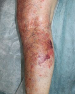
- The Dependent Risk Profile: Lesions follow a strict mechanical law, favoring dependent areas of the body lower than the heart where gravity causes blood to pool and immune complexes to settle more easily [cite: 1497, 1572, 1582]. Widespread, long-standing, or unmanaged lesions can progress to severe skin complications, including fluid-filled blisters (bullae), raw sloughing, or painful, deep cutaneous ulcers [cite: 1495, 1544, 1546].
Where and How It Appears on My Body
Vasculitis leaves a highly predictable geographical map across your body, displaying unique visual landmarks that guide your clinical team [cite: 1496, 1497, 1563].
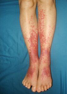
- The Dependent Landmark Anchor: The raised purple-red spots selectively target your lower extremities, clustering heavily around the ankles and lower legs [cite: 1497]. In extensive or highly active cases, the eruption can expand upward onto the thighs, buttocks, and occasionally the hands [cite: 627, 1497].
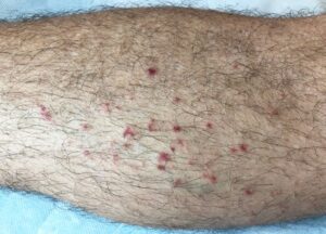
- The Visual Range: While classic palpable purpura appears as scattered, pinpoint-to-small purple bumps, a severe presentation can run together into massive, confluent purple-red plaques [cite: 1494, 1588, 1589]. Active areas can simultaneously display small blisters, pustules, or open sores [cite: 1495].
- The Non-Blanching Test: A cornerstone of identification is that purpuric spots are strictly fixed in color [cite: 1494, 1571]. When light pressure is applied to a spot, it does not blanch or fade to white; it remains a solid, deep purple because the blood is locked directly in the skin tissue rather than moving inside the vessel channel [cite: 1443, 1494].
- Muted Complexion Clues: On deeper skin tones, the early bright red or pink hue surrounding a spot can look muted, gray, or deeply hyperpigmented dark brown [cite: 1381, 1589]. In skin of color, tracing the distinct raised, bumpy texture of the purpura remains the primary clinical anchor used to locate active lesions [cite: 1494, 1571].
Solutions I Can Try at Home
Because cutaneous vasculitis is driven by an internal accumulation of immune complexes settled deep in your vessel walls, over-the-counter anti-itch lotions offer no structural benefit [cite: 1505, 1543]. At-home care focuses on mechanical strain reduction and tissue defense [cite: 1541, 1542].
- Enforce Strict Rest and Leg Elevation: This is your single most effective at-home healing tool [cite: 1541]. Keep your legs elevated above the level of your heart as frequently as possible during an active flare [cite: 1541]. Rest breaks gravity’s mechanical pulling force, immediately reducing localized lower leg fluid retention and easing throbbing leg pain or muscle aches [cite: 1541, 1578, 1582].
- Utilize Support Compression Hose: When you must stand or walk, wear appropriate support compression stockings on your lower limbs [cite: 1542]. Compression helps stabilize capillary filtration pressure and stops new immune complexes from settling easily into your dependent tissue layers [cite: 1542, 1572, 1582].
- Never Vigorously Scrub Active Spots: Avoid using rough loofahs, harsh washcloths, or coarse exfoliating scrubs on active purpuric areas. Because the vessel damage resides deep within the dermal layer, aggressive scrubbing splits the skin barrier lines, introduces surface bacteria, and can cause raw cutaneous ulcers to form [cite: 1495, 1544, 1569].
- Gently Log Exposure Timelines: Maintain a clear written record of your flare-up [cite: 1509]. Document exactly when the spots appeared and note if you started any new medications, changed your diet, or suffered from a sore throat or viral illness in the weeks preceding the rash, which provides your healthcare team with essential clues [cite: 1499, 1500, 1502, 1505].
When Should I See a Dermatology Provider?
Vasculitis is an unpredictable inflammatory condition that requires structured medical tracking [cite: 1510, 1567]. Seeking professional clinical triage early is important to execute specialized screenings, confirm your diagnosis, and implement prescription therapies before internal organs are affected [cite: 1509, 1510, 1567].
Seek Professional Help immediately if You Notice These Warning Signs:
- The Emergence of Widespread Purple Spots: You experience a sudden, rapid explosion of raised purple-red bumps or large confluent plaques across your legs, requiring urgent medical correlation [cite: 1494, 1588].
- Diagnostic Uncertainty: Your leg spots are expanding or changing, requiring an expert **office skin biopsy** (4mm punch) to confirm the structural presence of a neutrophilic vessel infiltrate and fragmented leukocytoclasia under a microscope [cite: 1509, 1565, 1570].
- The Development of Blisters or Open Sores: Active purpuric areas split open into large fluid-filled blisters (bullae) or raw, painful open ulcers, requiring immediate targeted medical wound management [cite: 1495, 1544].
- The Arrival of Internal or Systemic Flags: Your skin changes are accompanied by vital internal warning indicators, such as a persistent fever, sudden abdominal pain, blood in your urine or stool, a severe dry cough, or matching swelling and intense pain in multiple joints [cite: 1520, 1567, 1575, 1577].
Frequently Asked Questions
- Q: What extensive laboratory and diagnostic testing is mandatory to screen for underlying causes?
A: Because cutaneous vasculitis can be the initial external sign of an underlying autoimmune condition or internal organ disease, a positive diagnosis triggers a comprehensive baseline screening panel [cite: 1510, 1567]. Your provider will order targeted blood work, including a Complete Blood Count (CBC) with differential, a Comprehensive Metabolic Panel (CMP-14), an Antinuclear Antibody (ANA) test, and inflammatory markers (ESR/CRP) [cite: 1511, 1512, 1513, 1514]. To isolate the specific trigger, your team will explicitly run **anti-streptococcal antibodies, Hepatitis B and C serology, ANCA profiles, a formal Urinalysis to check your kidney filtration safety, and a serum protein electrophoresis with reflex to immunofixation (IFE) alongside cryoglobulins** to evaluate for abnormal immune complex configurations [cite: 1515, 1516, 1517, 1518, 1519, 1520, 1572]. - Q: What is the primary oral prescription protocol and taper schedule for progressive disease?
A: While mild, limited skin cases are often self-limiting and managed with local rest, severe, progressive, or ulcerating purpura requires systemic intervention [cite: 1539, 1546, 1547]. Your provider will initiate **oral Prednisone dosed at 0.5 mg/kg/day maintained strictly for 1 week** [cite: 1547]. Once your symptoms stabilize, your medical team will execute a highly structured, gradual **3-to-6 week steroid weaning taper** [cite: 1547]. Following this precise down-titration schedule is vital to safely minimize your risk of an intense, painful **inflammatory rebound flare** [cite: 1547]. For chronic, neutrophil-heavy skin configurations, oral **Dapsone at 50–100 mg daily** is an excellent alternative first-line step [cite: 1549]. However, **Dapsone is strictly contraindicated if you possess a G6PD enzyme deficiency** due to a life-threatening risk of severe red blood cell destruction (hemolysis) [cite: 1550, 1579]. Alternatively, oral azathioprine or methotrexate can be integrated [cite: 1552, 1553]. - Q: What advanced multi-step “CHAP Therapy” protocol exists for resistant or high-risk vascular disease?
A: For severe, chronic, or refractory configurations at a high risk for local tissue loss, your provider will step up care to the advanced **CHAP Therapy protocol** [cite: 1420, 1554]. Your dermatology team will prescribe these 4 highly targeted medications together to maximize blood vessel expansion, protect vascular linings, and keep your circulation moving cleanly [cite: 1421, 1554]:
- C – Calcium Channel Blockers: (Prescription **Nifedipine or Amlodipine**) to force direct, smooth vascular dilatation [cite: 1422, 1556].
- H – Hydroxychloroquine: To deliver targeted immunomodulation and soothe local lining inflammation pathways [cite: 1423, 1557].
- A – Aspirin: To safely decrease platelet aggregation and thin your blood flow [cite: 1424, 1558].
- P – Pentoxifylline: Dosed precisely at **400 mg three times a day** to lower your blood viscosity and actively prevent microvascular thrombosis formations from cutting off tissue oxygen [cite: 1425, 1559].
The long-term course of Cutaneous Small Vessel Vasculitis is typically favorable, with most isolated skin cases resolving completely on their own within a few weeks when managed with appropriate rest [cite: 1539, 1561]. Success relies on identifying and removing your specific environmental or drug trigger, maintaining strict leg elevation and compression guidelines during active attacks, and keeping up with your scheduled clinical follow-ups with your interprofessional care team to keep your body safe, healthy, and fully protected [cite: 1541, 1542, 1566].