
Sarcoidosis is a systemic inflammatory disease characterized by the formation of tiny clusters of immune cells called noncaseating granulomas [cite: 3450, 3458]. It is known in dermatology as "the great imitator" because its skin lesions can look like a wide variety of completely unrelated dermatological conditions [cite: 3402, 3451]. While skin bumps or patches are the initial sign of the disease in about one-third of individuals, sarcoidosis frequently involves internal organs, particularly the lungs and lymph nodes [cite: 3412, 3453]. Understanding your skin presentations through a professional biopsy and completing a thorough system-wide check ensures your body stays completely safe and healthy [cite: 3424, 3425, 3454].
What Is Causing My Skin Breakout?
Understanding how your body’s immune defenses react to your environment is a strategic first step in managing sarcoidosis. While the exact primary cause of sarcoidosis remains unknown, current medical evidence shows it is triggered by an abnormal, overactive immune response following exposure to an external environmental antigen in individuals with a genetic predisposition [cite: 3414]. These triggers can include everyday elements of both infectious and non-infectious origin, such as mold, mildew, insecticides, combustible wood, industrial dusts, or specific surface microbes [cite: 3415].
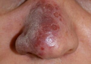
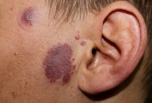
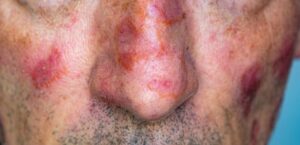
When an exposed individual encounters these antigens, it causes a dysregulated helper T-cell immune response [cite: 3416]. Your white blood cells launch an exaggerated defense, overproducing inflammatory signaling proteins (cytokines) like TNF-alpha, IL-12, IL-15, and IL-18 [cite: 3416, 3466, 3467]. Instead of clearing normally, these immune cells clump together tightly in your organs, forming microscopic structures called noncaseating granulomas [cite: 3416, 3458]. Identifying these cellular landmarks early is the essential “So What?” factor in your care plan [cite: 3454]. Because sarcoidosis is an internally driven systemic process, standard topical treatments alone cannot calm the broader immune signal, making expert screening and path-specific systemic therapies necessary to protect your tissue health [cite: 3425, 3433, 3439].
Understanding Specific vs. Non-Specific Skin Lesions
Sarcoidosis skin findings are divided strictly into two biological categories. Recognizing which type is present on your skin guides your medical testing and indicates your expected long-term course of healing [cite: 3403].
| Lesion Classification | Key Differentiators, Pathology, and Subtypes |
|---|---|
| Specific Lesions | Pathology: A skin biopsy explicitly reveals the presence of true noncaseating granulomas under the microscope [cite: 3404, 3470]. Major Variants: • Papular: Small raised bumps typically cluster across the face and are associated with a highly favorable outlook [cite: 3405, 3447]. • Plaque: Elevated, flat-topped patches populate the face, back, buttocks, and outer limbs, often indicating a higher risk of systemic organ involvement [cite: 3406, 3447]. • Lupus Pernio: Chronic, disfiguring, hardened reddish-purple areas line the nose, cheeks, and ears, strongly indicating severe internal disease [cite: 3407, 3473, 3474]. • Subcutaneous (Darier-Roussy): Deep, non-tender nodules settle under the skin of the extremities [cite: 3408]. • Scar Sarcoidosis: Granulomatous thickening develops directly within old, previously traumatized scars or injuries [cite: 3409]. |
| Non-Specific Lesions | Pathology: These represent a general reactive process; a skin biopsy shows localized tissue inflammation but lacks any internal granulomas [cite: 3410, 3471]. Primary Variant: • Erythema Nodosum (EN): Characterized by the sudden arrival of acute, highly painful, tender red nodules located predominantly across the shins [cite: 3410, 3475]. It typically carries an excellent prognosis, patterns resolution within 2 years, and rarely causes long-term scarring [cite: 3448, 3475]. |
Am I at risk for sarcoidosis or severe internal complications?
Developing sarcoidosis is an inflammatory event that can impact individuals of all backgrounds, though specific genetic templates and demographics dictate its overall severity [cite: 3414, 3417].
- The Gender and Racial Axis: Sarcoidosis demonstrates a statistically higher prevalence in women and Black individuals [cite: 3418]. The peak age of onset occurs during your thirties for the general population, shifting to your forties for African Americans [cite: 3419].
- Increased Severity Risk: Statistically, African American patients are far more likely to experience a highly severe, rapidly progressive disease course and demonstrate an increased risk of developing chronic, scarring cutaneous plaques compared to Caucasian individuals [cite: 3420].
- Extensive Internal Sparing Concerns: Because 90% to 95% of sarcoidosis patients experience internal pulmonary involvement, having a newly diagnosed specific skin lesion means you face a direct risk of hidden lung changes, requiring immediate baseline chest radiographs and function tests [cite: 3425, 3452].
- Chronic Fibrosis Trajectory: While granulomas resolve spontaneously within 2 to 5 years in 60% of cases, approximately 20% to 30% of individuals progress to permanent tissue scarring (fibrosis), locking them into a chronic disease course [cite: 3445, 3446, 3468, 3469].
Where and How It Appears on My Body
Sarcoidosis lesions leave distinct physical “maps” and visual landmarks across your body that require careful clinical identification [cite: 3411, 3454].
- The Facial Mask: Specific papular lesions and disfiguring lupus pernio plaques group heavily around the central facial triangle—predominantly lining the contours of the nose, eyelids, and cheeks [cite: 3405, 3407, 3473].
- The Extensor Distribution: Chronic plaque formations show a strong preference for the upper back, buttocks, and the extensor surfaces of the elbows and knees [cite: 3406].
- The “Apple Jelly” Hue (Diascopy Landmark): When a dermatology provider presses a clear glass slide firmly against a reddish-brown sarcoidosis plaque (a clinical test called diascopy), the background redness is safely blanched away, revealing a highly unique, translucent yellowish-brown “apple jelly” hue underneath [cite: 425].
- Muted Complexion Trajectories: On deeper skin tones, the classic reddish-purple color of specific papules can appear muted, deep brown, or hypopigmented (lighter than surrounding skin) [cite: 3407, 3431]. In dark skin, reactive erythema nodosum on the shins may mimic deep hyperpigmentation rather than bright red spots [cite: 1886].
Solutions I Can Try at Home
Because moderate-to-widespread cutaneous sarcoidosis is driven by an internal, systemic inflammatory highway, at-home care focuses entirely on skin barrier protection, logging symptoms, and supporting tissue health [cite: 3416, 3433].
- Track Border Changes Safely: Maintain a clear photographic log of your skin lesions. Documenting whether plaque borders stay stable or slowly expand over several weeks provides invaluable objective evidence for your interprofessional medical team [cite: 3433].
- Do Not Pick or Scratch Scars: If your sarcoidosis populates old scar lines, leave them completely alone [cite: 3409]. Aggressively picking or scratching at the skin breaks your protective barrier, intensifies local cytokine release, and risks introducing a secondary bacterial infection [cite: 3416, 500].
- Lubricate and Protect: Keep your general skin heavily moisturized with thick, fragrance-free emollient creams to reduce localized surface flaking and keep your skin surface resilient against external friction [cite: 2691].
When Should I See a Dermatology Provider?
Sarcoidosis can easily mask itself as common tinea, psoriasis, or lupus [cite: 3431]. Seeking professional clinical triage early is vital to get a precise diagnosis through an office **4mm punch skin biopsy** to check your tissue histology for noncaseating granulomas and rule out dangerous lookup mimics like cutaneous T-cell lymphoma or infectious tuberculosis [cite: 3424, 3431, 3432].
Seek Professional Help immediately if You Notice These Warning Signs:
- The “First Plaque” or Changing Nodule: You notice a new, expanding, or changing reddish-brown plaque that has completely failed to respond to basic over-the-counter creams [cite: 3431, 3432].
- Disfiguring Facial Hardening: Hardened, purple, or highly visible vascular plaques begin developing across your nose, cheeks, or ears, indicating lupus pernio and requiring urgent intervention to halt cosmetic damage [cite: 3407, 3473].
- New Internal Organ Signaling: Your skin changes are accompanied by pulmonary or systemic warning flags, such as a persistent dry cough, unexplained shortness of breath, sudden chest pain, eye vision shifts, or an irregular heartbeat (arrhythmia) [cite: 3425, 3427, 3443].
Frequently Asked Questions
- Q: What extensive internal medical tests must be executed if my skin biopsy is positive for sarcoidosis?
A: Because sarcoidosis features an exceptionally high rate of internal pulmonary involvement, a positive skin biopsy automatically triggers a comprehensive diagnostic screening protocol [cite: 3424, 3425]. Your interprofessional medical team will order a posteroanterior chest radiograph (CXR) and formal pulmonary function tests (PFTs) to map your lungs [cite: 3427, 3443]. This is paired with an electrocardiogram (EKG) and echocardiogram to check your heart, a formal ophthalmologic slit-lamp examination to screen for hidden eye inflammation (uveitis), and a tuberculin skin test or IGRA to rule out tuberculosis before starting treatment [cite: 3427, 3443]. Your baseline blood panel will actively track complete blood counts, renal function (CMP), serum ACE levels, and vitamin D markers [cite: 3426, 3427]. - Q: What primary medical treatments and steroid-sparing medications will my provider prescribe?
A: For limited, mild skin lesions, high-potency Class I topical steroid creams or direct intralesional corticosteroid injections are used as first-line options to calm localized granulomas [cite: 3436, 3482]. However, if your skin lesions are widespread, disfiguring, or involve internal organs, your provider will initiate systemic corticosteroids [cite: 3439]. To limit the long-term side effects of steroids, your team will integrate powerful steroid-sparing agents, most commonly anti-malarials like **hydroxychloroquine or chloroquine** (exceptionally effective for facial lupus pernio) or weekly **methotrexate** [cite: 3437, 3438, 3439, 3482, 3483]. For severe, refractory lesions that fail standard care, advanced anti-TNF alpha biological therapies (such as infliximab or adalimumab) can be safely added to your regimen [cite: 3440, 3484]. - Q: How often must I follow up with my medical team to ensure my safety?
A: Sarcoidosis requires structured, ongoing clinical tracking [cite: 3443]. If you are actively undergoing therapy or taking systemic medications, you must return for a formal evaluation **every 6 months** [cite: 3442]. If your condition becomes completely stable and you are off active medications, your follow-up schedule transitions to an **annual checkup** [cite: 3442]. Each regular evaluation includes a thorough physical exam alongside repeat safety screenings, including an annual eye exam, EKG, chest radiograph, lung function tracking, and complete serum chemistries to confirm your body remains fully protected [cite: 3443].
The long-term outlook for the majority of sarcoidosis patients is highly favorable, with skin lesions and granulomatous inflammation clearing completely on their own within 5 years in most cases [cite: 3445]. Success relies on swift clinicoradiographic verification via an office biopsy, long-term commitment to your systemic monitoring schedule, and an enduring collaborative partnership with your interprofessional healthcare team to keep your body vibrant and healthy [cite: 3424, 3442, 3443, 3454].