Quick Summary: Pyoderma Gangrenosum (PG) is a rare, non-infectious inflammatory skin disorder characterized by the rapid and painful development of skin ulcers [cite: 1562, 1620, 1626]. Classified as a neutrophilic dermatosis, it is often associated with an underlying systemic condition [cite: 1572, 1574, 1622]. Early clinical staging and specialized management are critical milestones to halt tissue destruction, minimize scarring, and safely restore wound healing [cite: 1599, 1616, 1623].
What Is Causing These Painful Skin Ulcers?
Understanding how your body’s immune defense mechanisms interact with your skin layers is a strategic first step in managing Pyoderma Gangrenosum. Despite its name, PG is completely non-infectious and is an idiopathic condition [cite: 1570, 1620, 1627]. It is fundamentally an autoinflammatory disease where the body’s immune system mistakenly attacks healthy skin tissue [cite: 1571, 1629].
The underlying disease process identifies PG as one of the neutrophilic dermatoses [cite: 1572, 1635]. This means it is characterized by an overactive, excessive accumulation of specific white blood cells—called neutrophils—acting in response to an internal antigen [cite: 1572, 1635]. You can think of an active PG ulcer as an internal “immune wildfire.” When these overactive white blood cells flood the skin tissue, they cause rapid cell breakdown and deep tissue destruction [cite: 1562, 1563, 1620].
Identifying this condition early through an expert physical examination is the essential “So What?” factor in your recovery plan [cite: 1581, 1582, 1616]. Because PG operates via deep autoinflammatory pathways rather than a simple surface allergen or bacterial infection, traditional over-the-counter antibiotic ointments are completely ineffective, making targeted anti-inflammatory and systemic immune therapies mandatory to protect your tissue lines [cite: 1599, 1601, 1609, 1620].
Understanding the Condition: Comparing PG to Sweet’s Syndrome
Pyoderma Gangrenosum belongs to the same broad family of neutrophilic dermatoses as another rare inflammatory skin condition called Sweet’s syndrome [cite: 1572, 1596, 1635]. Recognizing the physical and structural variations between these conditions prevents medical confusion and directs your provider toward the right therapeutic approach [cite: 1581, 1590].
| Neutrophilic Condition | Key Differentiators and Visual Configurations |
|---|---|
| Sweet’s Syndrome | Appearance: Presents as raised, solid pink-to-red bumps, nodules, or plaques [cite: 1596, 1633]. These lesions typically remain solid and lack rapid, deep tissue ulceration [cite: 1562, 1596]. It commonly affects the face, neck, or hands and is classically accompanied by systemic signs like acute fever, joint pain (arthralgia), and malaise [cite: 1596, 1597, 1636, 1637]. |
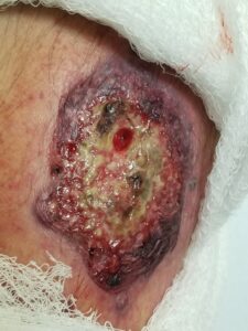
| Pyoderma Gangrenosum (PG) | Appearance: Characterized by rapidly expanding, deep skin ulcers [cite: 1562, 1626, 1631]. Lesions quickly erode from small bumps or pus-filled blisters into open sores featuring a necrotic base and a distinct, raised, overhanging border with a characteristic blue or purple “undermined” edge [cite: 1563, 1564, 1565, 1632]. |
Am I at risk for Pyoderma Gangrenosum or severe complications?
Developing Pyoderma Gangrenosum is an autoinflammatory medical event [cite: 1571, 1621]. It has absolutely zero connection to poor personal hygiene, personal cleanliness flaws, or contagious environmental exposures [cite: 1620, 1627].
- Systemic Comorbidity Links: Approximately half of all documented PG cases are directly associated with an underlying systemic health condition [cite: 1574, 1646]. Major risk associations include inflammatory bowel diseases (such as ulcerative colitis and Crohn’s disease), rheumatoid arthritis, and underlying blood disorders (including leukemia and lymphoma) [cite: 1575, 1576, 1577, 1646].
- The Pathergy Phenomenon: Your skin cells maintain a highly specialized, hyper-reactive trait known as **pathergy** [cite: 1579, 1630]. This means that minor skin trauma, scratches, cuts, needle pricks, or even formal surgical incisions can act as a direct trigger to launch or severely worsen a rapidly expanding PG ulcer right at the injury site [cite: 1579, 1607, 1630].
- Peristomal Stoma Risks: Individuals who have undergone abdominal surgeries requiring an artificial opening are at risk for a specialized presentation known as **peristomal pyoderma gangrenosum**, where painful ulcers rapidly form directly in the skin surrounding the stoma site [cite: 1568, 1644].
- The Scarring Trajectory: Because PG ulcers cause significant and deep tissue destruction, healing is a slow process that frequently leaves behind long-lasting, distinct scars that can feature a unique, criss-cross pattern known clinically as **cribriform scarring** [cite: 1565, 1615, 1616, 1649].
Where and How It Appears on My Body
Pyoderma Gangrenosum leaves an unmistakable physical map across your skin, displaying unique visual landmarks that help your dermatology provider distinguish it from standard vascular or pressure ulcers [cite: 1564, 1581, 1594].
- The Lower Extremity Preference: Although PG can arise anywhere on the body, ulcers show a massive, overwhelming biological preference for the lower extremities—clustering heavily across the legs, ankles, and feet [cite: 1567]. Secondary sites include the arms, genitals, and neck lines [cite: 1568].
- The Undermined Purple Edge: Tracing the margin of an active ulcer reveals a highly unique overhanging or “undermined” edge [cite: 1564]. This border is typically raised, slightly rolled, and displays a prominent blue, dusky violaceous, or purple halo that marks the line of active inflammation expanding into healthy tissue [cite: 1564, 1592, 1626].
- The Soft, Boggy Core: Unlike many chronic ulcers that feel hard and indurated at the borders, the active edges of a PG wound are characteristically soft, boggy, and pliable to the touch [cite: 1593]. The base of the open sore often holds a necrotic or purulent fluid discharge [cite: 1563].
- Muted Complexion Clues: On deeper skin tones, the classic violaceous or purple hue of the undermined border can look muted, dark brown, or gray, blending into deep post-inflammatory hyperpigmentation [cite: 1564, 1650]. On skin of color, evaluating localized skin tenderness and watching for a soft, boggy texture are vital clinical anchors used to spot active changes [cite: 1565, 1593].
Solutions I Can Try at Home
Because Pyoderma Gangrenosum is a deep, internally driven autoinflammatory process, home remedies provide zero benefit [cite: 1571, 1620]. At-home support focuses entirely on protecting your skin from accidental trauma to avoid fueling the pathergy cycle [cite: 1579, 1618, 1630].
- Meticulously Protect Skin from Trauma: Avoid all accidental cuts, scratches, or bumps to your skin, especially on your lower legs [cite: 1618]. Because of the pathergy effect, a simple minor scratch can rapidly evolve into a large, painful open ulcer [cite: 1563, 1579, 1630]. Wear soft, protective clothing and avoid harsh physical environments [cite: 1618].
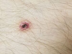
- Do Not Pick or Squeeze Bumps: If you notice a small, discolored bump or a new pus-filled tip, leave it completely alone [cite: 1563, 1632]. Squeezing or popping a lesion manually acts as a mechanical trauma that pushes the neutrophilic infiltrate deeper into your tissue and triggers rapid ulceration [cite: 1563, 1579, 1643].
- Execute Gentle Wound Care: Keep your existing ulcers covered with specialized clean dressings according to your provider’s explicit wound protocol [cite: 1606]. Maintaining a clean, protected environment supporting moist wound healing helps insulate the thinned, fragile tissue edges from external friction [cite: 1606, 1650].
When Should I See a Dermatology Provider?
Pyoderma Gangrenosum is an unpredictable medical condition that can progress with alarming speed [cite: 1562, 1615]. Seeking professional clinical triage early is vital to rule out dangerous lookalikes and secure a path-specific treatment plan [cite: 1581, 1584, 1616, 1623].
Seek Professional Help immediately if You Notice These Warning Signs:
- Rapidly Expanding Open Sores: A small skin bump or pustule quickly breaks open and turns into an intensely painful, expanding ulcer over a matter of days [cite: 1562, 1563, 1565].
- Diagnostic Uncertainty: You are struggling with non-healing leg ulcers that mimic common venous stasis or arterial disease, requiring your provider to perform a diagnostic **office skin biopsy** [cite: 1581, 1585, 1594]. Pathologists utilize a specialized, large **5-6 mm punch biopsy** taken specifically from the overhanging, overyielding edge of the ulcer to capture enough deep tissue architecture to support a definitive diagnosis of PG [cite: 1586, 1587].
- Signs of a Secondary Superinfection: The ulcerated tissue becomes exquisitely tender, develops a foul odor, or drains thick yellow pus, suggesting a secondary infection that requires immediate tissue culture swabs to separate PG inflammation from a live bacterial invasion [cite: 1591, 1620].
- Systemic Comorbidity Warnings: Widespread or painful skin breakages are accompanied by unprompted weight loss, recurrent chronic diarrhea, severe abdominal pain, or blood in your stool, suggesting an undiagnosed inflammatory bowel disease or hematological shift [cite: 1575, 1577, 1589].
Frequently Asked Questions
- Q: What extensive multi-system screening is executed to search for underlying PG triggers?
A: Because approximately 50% of Pyoderma Gangrenosum cases stem from internal conditions, a positive clinical staging automatically initiates a thorough medical evaluation [cite: 1574, 1581, 1646]. Your interprofessional healthcare team will order comprehensive blood work, including a Complete Blood Count (CBC) with differential to screen for leukemia or lymphoma, a Comprehensive Metabolic Panel (CMP), and inflammatory markers [cite: 1585, 1589, 1643]. Depending on your history, your team will schedule a formal rectal examination, bone marrow sampling, or an urgent **screening colonoscopy** to explicitly evaluate your GI tract for hidden ulcerative colitis or Crohn’s disease [cite: 1575, 1589]. A pathergy skin prick test can also be incorporated [cite: 1588]. - Q: What highly specialized topical and intralesional medication protocols are available?
A: For limited, mild-to-moderate areas of disease, your provider will focus on calming the active borders using targeted local anti-inflammatory therapies [cite: 1600, 1601]. First-line choices include the application of superpotent Topical Class I or Class II corticosteroid creams directly to the ulcer margins [cite: 1601]. Alternatively, your provider can compound a highly effective **Topical Cyclosporine Solution** right in the office, created precisely by mixing **two 100mg capsules of Cyclosporine with 98ml of Vitamin E oil** to apply once daily (QD) or twice daily (BID) to promote wound healing [cite: 1599, 1603]. For targeted core suppression, your provider can use **one ml of cyclosporine eye drops (Restasis 0.05%) applied twice daily** directly onto the active ulcer bed, or perform precise **intralesional steroid injections** straight into the overhanging ulcer edge [cite: 1604, 1605]. - Q: What advanced oral systemic options and surgical rules exist for severe, refractory disease?
A: For severe, widespread, or rapidly progressive variants that fail local measures, your provider will step up care to internal treatments [cite: 1609, 1615, 1617]. Systemic interventions include anti-inflammatory tetracycline antibiotics, or oral **Dapsone**, which is highly valued for its unique ability to safely suppress overactive neutrophils [cite: 1610, 1611, 1635]. Advanced immunosuppressants like oral cyclosporine, azathioprine, mycophenolate, or modern oral JAK inhibitors can also be integrated into your treatment algorithm [cite: 1612, 1613, 1639]. Crucially, if you require surgery or surgical debridement of dead tissue, it must be executed with extreme caution due to the pathergy effect [cite: 1607, 1630]. To shield your skin, patients with a documented history of PG will receive **prophylactic systemic corticosteroids prior to any surgery** to stabilize their immune baseline and actively prevent a life-threatening recurrence [cite: 1608, 1617].
The long-term course of pyoderma gangrenosum can be highly unpredictable, with ulcers occasionally requiring weeks or multiple months to achieve complete closure [cite: 1615]. While the condition is complex, early diagnostic correlation combined with modern targeted local or systemic autoinflammatory therapies can successfully calm your immune signaling, control pain, and promote clean wound healing [cite: 1599, 1616, 1623]. Success relies on absolute avoidance of skin trauma, strict adherence to your compounded or morning medication routine, and maintaining a regular relationship with your interprofessional dermatology team to keep your body safe and fully supported [cite: 1581, 1618, 1624].