Pityriasis lichenoides (PL) is an uncommon, chronic inflammatory skin condition characterized by recurring crops of small bumps (papules) that can appear across various parts of the body. This condition exists on a fluid disease spectrum and presents in two primary clinical forms: an acute type known as Pityriasis Lichenoides et Varioliformis Acuta (PLEVA) and a chronic type called Pityriasis Lichenoides Chronica (PLC). While the sudden appearance of these recurring crops can look concerning, the condition is generally benign and self-limiting. Successful long-term management relies on obtaining an accurate diagnosis through a professional skin biopsy, tailored anti-inflammatory therapies, and consistent tracking of skin changes.
What Is Causing These Recurring Bumps?
Understanding how your body’s immune pathways respond to external and internal triggers is a strategic first step in managing pityriasis lichenoides. While the exact cause of this condition remains unknown, scientific hypotheses suggest it represents an inflammatory hypersensitivity reaction. You can think of a flare-up as an “overactive immune defense response”—where your white blood cells (T-lymphocytes) multiply and gather persistently, targeting the junction where your outer skin layer meets the deeper tissue.
This localized immune response is frequently initiated by common infectious triggers or external exposures. Associated triggers include:
- Viral Agents: Epstein-Barr Virus (EBV), cytomegalovirus (CMV), and HIV.
- Bacterial Microbes: Staphylococcus and Streptococcus strains.
- Parasitic Organisms: Toxoplasma gondii.
- Medication Profiles: Certain prescription therapies, including anti-TNF agents, statins, antidepressants, vaccines, and radiocontrast dyes.
Identifying this condition early is the essential “So What?” factor in your recovery plan. Because it is a low-grade immune-mediated process rather than a simple surface allergy, establishing a precise diagnosis prevents medical guesswork and guides your clinician toward targeted anti-inflammatory regimens.
Understanding the Disease Spectrum: PLEVA vs. PLC
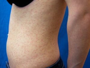
Pityriasis lichenoides operates as a fluid disease spectrum. An active case can present as a sudden, intense eruption (PLEVA), exhibit a slow, long-term flaking pattern (PLC), or visually transition from the acute phase into the chronic form over time.
| Disease Phase | Key Differentiators and Visual Profiles |
|---|---|
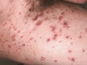
PLEVA (Acute Phase) | Appearance: Characterized by the abrupt arrival of crops of reddish-brown, oval bumps. These rapidly evolve into fluid-filled blisters (vesicles), small pus-filled tips (pustules), dark bloody scabs (hemorrhagic crusts), and shallow open ulcers. Sensation: Typically asymptomatic, though lesions can cause mild local itching or a sharp burning quality. |
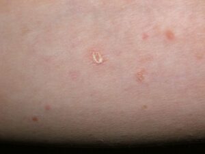
PLC (Chronic Phase) | Appearance: Characterized by smaller, dry, reddish-brown or brown-hued bumps. Active spots display a highly unique, adherent, shiny, and wafer-like “mica-like” scale that becomes distinctly more prominent and visible when scratched. Spots present simultaneously in completely different stages of evolution. Sensation: Asymptomatic or accompanied by mild, fluctuating surface itching. |
Am I at risk for pityriasis lichenoides?
Developing pityriasis lichenoides is an isolated biological event. It has absolutely zero connection to personal hygiene, dietary choices, skin cleanliness, or a globally compromised immune system.
- The Youth Profile: While it can populate any age group, it demonstrates a clear statistical preference for children, adolescents, and young adults under 30 years of age.
- Gender Demographics: It affects individuals across all races, showing a mild demographic preference for males over females.
- The Clinical Spectrum Risk: Having an active primary case of PLEVA places you at a direct risk for long-term skin changes, as the acute lesions frequently flatten out and evolve directly into chronic, slow-clearing PLC spots.
- Systemic Health Connections: Individuals recovering from a recent bacterial or viral illness (such as strep throat or mono) may experience an increased risk of triggering this reactive dermatosis.
Where and How It Appears on My Body
Pityriasis lichenoides leaves a specific physical “map” of landmarks across the body surface that helps your dermatology provider easily separate it from standard drug allergies or chickenpox.
- Trunk and Proximal Extremities: Lesions show a profound preference for the torso, abdomen, chest, upper arms, and thighs.
- Anatomic Sparing: Unlike viral chickenpox, it is very uncommon for classic pityriasis lichenoides to involve or spread extensively across the face, scalp, palms, or soles of the feet.
- Crops in Varying Stages: In chronic PLC, old spots will flatten into brown stains while brand-new scaly bumps emerge right next to them, creating a mixed, multi-stage visual layout.
- Muted Color and Shadows: On deeper skin complexions, the classic reddish-brown color of the papules can look muted, deep brown, or purplish. As active spots resolve, they commonly leave behind long-lasting shadows of light skin (postinflammatory hypopigmentation) or dark marks, which naturally blend back into your baseline skin tone over time.
Solutions I Can Try at Home
Because pityriasis lichenoides is an internally driven skin condition, home care focuses heavily on soothing active skin irritation, protecting the tissue barrier, and preventing scarring.
- Avoid Scratching Wafer Scales: Do not aggressively pick, scratch, or attempt to peel off the thick, mica-like wafer scales. Scratching active spots triggers further local cell irritation, prolongs the overall healing timeline, and risks introducing a secondary bacterial infection.
- Keep Skin Cool and Soothed: Apply lightweight, fragrance-free moisturizing creams or bland emollients regularly to alleviate surface flaking. Taking short, lukewarm showers and avoiding high-heat environments helps quiet localized nerve endings and reduces itch signals.
- Track Eruptive Crops: Take clear, well-lit photographs of identical skin regions on your arms or torso every two weeks. Maintaining a structured visual diary provides your clinical team with objective evidence to evaluate whether your spots are successfully flattening or expanding.
When Should I See a Dermatology Provider?
A professional evaluation is critical for pityriasis lichenoides. Because early lesions can look identical to common eczema or an allergic drug rash, your provider must perform a safe, in-office 4mm punch skin biopsy to look at the tissue structure under a microscope and rule out more serious lookalikes.
Seek Professional Help if You Notice These Warning Signs:
- Diagnostic Uncertainty: You have a persistent, widespread rash that has failed to improve with standard over-the-counter moisturizers or anti-itch creams.
- The Multi-Month Extension: Active scaly patches or recurring crops of bumps fail to flatten out or continue to spread dynamically past 3 to 4 months, requiring careful clinicopathological correlation to rule out conditions like cutaneous T-cell lymphoma.
- Severe Skin Breakdown: Active PLEVA spots turn into highly painful, open, or fluid-draining ulcers, or develop spreading black crusted sores that suggest a severe, ulceronecrotic progression.
- Signs of a Secondary Infection: You notice localized heat, red streaks climbing up a limb, or honey-colored crusting, indicating bacteria have invaded a scratched lesion.
Frequently Asked Questions
- Q: What primary medical treatments will my provider prescribe to clear the rash?
A: If your condition is extensive or causing discomfort, your provider will prescribe targeted first-line therapies. A cornerstone of management is a course of oral antibiotics, such as erythromycin or doxycycline; these are chosen specifically for their powerful, internal anti-inflammatory mechanisms rather than to fight a live infection. This is routinely paired with topical corticosteroid creams or non-steroidal topical immunomodulators (like tacrolimus or pimecrolimus) to calm surface irritation safely. - Q: What advanced treatment options exist for severe or highly resistant cases?
A: For severe, chronic, or recalcitrant cases that do not respond fully to first-line oral antibiotics and topicals, your dermatologist can step up therapy safely. In-office phototherapy sessions utilizing narrowband UVB or PUVA light can be exceptionally effective at slowing down overactive skin cells and clearing extensive plaques. For globally widespread or scarring disease, low-dose weekly regimens of systemic medications like methotrexate (MTX) can be incorporated under close medical supervision. - Q: Is pityriasis lichenoides contagious, and can it turn into skin cancer?
A: No, pityriasis lichenoides is entirely non-contagious; it cannot be caught from or spread to anyone else through physical contact or shared objects. Structurally, it is an inflammatory condition that runs a benign, self-limiting course over months to a few years. However, because rare instances of malignant transformation into lymphoma have been documented in scientific literature, maintaining a regular follow-up schedule with your dermatology team is essential to ensure long-term safety.
The long-term outlook for pityriasis lichenoides is highly favorable, with the vast majority of healthy patients experiencing a complete, natural clearing of the spectrum without long-term health issues. Success relies on early diagnostic confirmation via an office biopsy, long-term consistency with your anti-inflammatory routine, and close, ongoing partnership with your dermatology provider to track your recovery smoothly.