Necrobiosis Lipoidica (NL) is a rare, chronic granulomatous skin disorder that characteristically presents as distinct yellow, orange, or reddish-brown thinned patches on the skin. While these patches can develop anywhere on the body, they demonstrate a profound biological preference for the lower legs. NL is a complex, long-standing condition that is heavily linked to underlying metabolic health and blood vessel stability. Understanding your skin presentations through a professional biopsy, protecting your skin from physical injury, and establishing a tailored management plan are vital steps to minimize tissue damage, control active inflammation, and protect your long-term health.
What Is Causing These Discolored Lower Leg Patches?
Understanding how small blood vessels and immune pathways interact within your skin layers is a strategic first step in managing necrobiosis lipoidica. The exact root cause of NL remains unknown. However, strong clinical evidence points to a process called microvascular damage—where chronic, low-grade injury to your skin’s smallest blood vessels compromises the normal blood supply to the tissue. This localized lack of proper blood flow triggers abnormal collagen degeneration and a secondary, overactive inflammatory response deep within the dermis.
When this internal cascade is triggered, your white blood cells—specifically histiocytes, giant cells, and lymphocytes—gather persistently to wall off the damaged collagen fibers, creating microscopic structures known as granulomas. You can think of an NL patch as an area of internal “microvascular architectural failure.” Over time, the loss of blood flow causes the outer skin layer to thin out and clear, making the deeper, widened blood vessels underneath highly visible on the surface. Identifying these changes early through a professional clinical evaluation is the essential “So What?” factor in your care, providing you with an early warning signal to evaluate your broader metabolic health and implement targeted therapies before the skin becomes fragile or breaks open.
Understanding the Condition: Comparing NL to Granuloma Annulare
Necrobiosis lipoidica belongs to the same broad family of granulomatous disorders as another common skin condition called Granuloma Annulare (GA). Recognizing the key physical and architectural differences between these two lookup mimics prevents medical confusion and guides your clinician toward the correct therapeutic path.
| Skin Condition | Key Differentiators and Visual Configurations |
|---|---|
| Granuloma Annulare (GA) | Presents as firm, smooth, non-scaling pink or skin-colored bumps arranged in a neat, circular ring. The skin in the very center of the ring appears completely normal and healthy, lacking any internal thinning, yellow discoloration, or prominent visible surface blood vessels. |
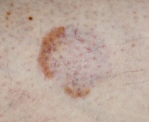
| Necrobiosis Lipoidica (NL) | Forms flat, irregular, yellow-orange or reddish-brown plaques. The central portion is highly shiny, porcelain-like, and thinned out (atrophic), showing an extensive network of tiny, visible spider-like blood vessels known as telangiectasias. |
Am I at risk for Necrobiosis Lipoidica or diabetes complications?
Developing necrobiosis lipoidica is an isolated vascular and inflammatory event. It has absolutely zero connection to personal hygiene flaws, skin cleanliness, or a globally deficient immune system.
- The Diabetes Nexus: The single most notable and powerful risk factor is diabetes mellitus. While it can technically surface in non-diabetic individuals, the presence of NL serves as a strong clinical marker for underlying glucose intolerance, a family history of diabetes, or progressive microvascular end-organ damage.
- The Gender Axis: NL demonstrates a profound biological preference for women, predominantly targeting females between their mid-20s and mid-40s.
- The Tobacco Multiplier: Smoking acts as a massive primary biological accelerator for this disease. Tobacco smoke directly damages blood vessel linings, severely worsening localized microangiopathy and increasing your risk for severe skin flares.
- Other Systemic Links: Susceptibility is mildly influenced by a cluster of secondary metabolic conditions, including thyroid disorders, hypertension, truncal obesity, and dyslipidemias (abnormal cholesterol levels).
Where and How It Appears on My Body
Necrobiosis lipoidica leaves a highly specific anatomical “map” across your limbs, presenting unique visual hallmarks that help your dermatology provider instantly identify it.
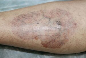
- The Anterior Shin Anchor: Lesions show a massive, overwhelming preference for the anterior shins (the front of the lower legs). They typically pop up symmetrically on both legs simultaneously.
- The Atrophic Glaze and Telangiectasias: Established plaques take on an irregular shape with a firm, reddish-brown or purple outer border, while the center transforms into a smooth, thin, yellow-orange glaze. Looking closely reveals prominent, winding, tree-like telangiectatic vessels branching across the central aspect.
- The Traumatic Ulceration Risk: Because the thinned skin over the shin bone has a compromised blood supply, it is exceptionally fragile. Up to 35% of patients develop deep, slow-healing, and incredibly painful skin breakages (ulcerations) within the center of the plaques, often triggered by a minor, accidental bump to the leg.
- Muted Complexion Clues: On deeper skin tones, the classic yellow-orange central hue may appear muted, dusky brown, or darkly hyperpigmented. In skin of color, the outer border often displays a deep purple or violaceous ring that remains elevated against the thinned center.
Solutions I Can Try at Home
Because necrobiosis lipoidica is a deep-seated, chronic granulomatous condition linked to small vessel damage, home care focuses on protecting your legs from physical trauma and eliminating external vascular stressors.
- Commit to Immediate Smoking Cessation: This is the single most vital behavioral tool you can implement. Stopping smoking removes a powerful toxin that constricts your small blood vessels, helping to stabilize your local tissue repair function.
- Meticulously Protect Your Shins: Wear thick, protective socks, shin guards during physical activities, or long trousers to insulate your lower legs from accidental contact. Preventing localized skin trauma actively stops the rapid, linear tracking of new lesions, a process known as koebnerization.
- Optimize Your Glycemic Control: Work closely with your primary care provider or endocrinologist to stabilize your blood sugar levels and track your Hemoglobin A1c (HbA1c). While optimizing diabetes control does not always automatically reverse existing skin plaques, it supports global microvascular health and prevents further end-organ damage.
- Do Not Scrub or Exfoliate: Never try to aggressively rub active plaques or pick at the central thinned skin. Scrubbing the skin will split the fragile tissue lines open, causing painful ulcerations that are exceptionally difficult to heal.
When Should I See a Dermatology Provider?
NL is a chronic condition that requires careful medical monitoring. Because early papules can look identical to common stasis dermatitis, sarcoidosis, or deep fungal infections, your provider must perform a safe, in-office **4mm punch skin biopsy** to evaluate the dermal layers under a microscope and rule out dangerous lookup mimics.
Seek Professional Help immediately if You Notice These Warning Signs:
- The Development of Open Ulcers: The thinned center of your shin plaque splits open, begins weeping fluid, or forms a raw, painful sore (ulceration), requiring immediate specialized wound care.
- Worsening Pain or Altered Sensation: An asymptomatic plaque suddenly becomes highly painful, develops an unyielding burning sensation (dysesthesia), or loses feeling entirely (anesthetic skin), indicating progressive nerve and tissue injury.
- Rapid Peripheral Expansion: Your leg patches expand rapidly or multiply across your extremities, causing significant cosmetic distress or local swelling.
- Signs of a Secondary Infection: An open leg sore becomes intensely hot, swollen, increasingly red, or drains foul-smelling pus, suggesting a bacterial superinfection or deep bone infection (osteomyelitis) requiring urgent systemic antibiotics.
Frequently Asked Questions
- Q: What primary medical treatments will my provider use to control active NL inflammation?
A: Because NL is an internally driven dermal process, treatment can be challenging. First-line clinical interventions focus on calming the active inflammatory borders. Your provider will prescribe super-high-potency **topical corticosteroid creams**, which are often applied under plastic wrap or a specialized dressing (occlusion) to maximize absorption into the deep tissue. For stubborn or expanding plaques, your dermatologist can perform in-office **intralesional corticosteroid injections** directly into the active reddish-brown margins to rapidly halt local granuloma formation. Alternative local options include topical tacrolimus or topical retinoids. - Q: What advanced systemic treatments exist for severe, recalcitrant, or ulcerated cases?
A: For severe, rapidly progressive, or ulcerated variants that fail standard topicals, your provider will step up care to systemic interventions. Advanced therapies include **PUVA photochemotherapy** or oral antimalarials like hydroxychloroquine. Emerging medical evidence also demonstrates high success rates utilizing targeted **biologic agents (TNF inhibitors)** to safely block the pro-inflammatory cytokines driving the disease and accelerate wound closure. If a severe ulcer fails to respond to all medical therapies, surgical wide excision combined with **split-thickness skin grafting** can be performed as an advanced rescue option. - Q: Why is a skin biopsy necessary, and how does it separate NL from serious lookalikes?
A: A skin biopsy is critical because NL shares a virtually identical color palette and shin distribution with multiple serious conditions. Most notably, a biopsy is executed to distinguish NL from **necrobiotic xanthogranuloma**, a severe skin marker that serves as a major red flag for underlying malignant plasma cell dyscrasias and paraproteinemia. Pathologists can cleanly separate these conditions under a microscope by evaluating your tissue layers. While necrobiosis lipoidica explicitly displays layered, tiered granulomatous inflammation of histiocytes and giant cells running parallel to degenerating dermal collagen, other mimics show completely different cellular signatures, providing you with absolute diagnostic certainty.
The long-term course of necrobiosis lipoidica is typically chronic, requiring ongoing patience and regular clinical monitoring. While spontaneous remission is rare, modern targeted therapies can successfully stabilize active borders, support your skin’s structural resilience, and protect your lower legs from complications. Success relies on absolute tobacco avoidance, strict adherence to shin protection protocols, and maintaining a close, collaborative relationship with your interprofessional dermatology team to keep your body healthy and fully supported.