Granuloma Annulare (GA) is a common, chronic inflammatory skin condition characterized by distinctive ring-shaped (annular), smooth bumps and plaques [cite: 1074, 1121]. While the appearance of these expanding rings can cause concern, GA is a completely benign condition that is not a form of skin cancer and is not contagious [cite: 1083, 1121]. In many cases, the condition is entirely self-limiting and will disappear naturally on its own over time [cite: 1109, 1123]. Clinical management focuses on confirming an accurate diagnosis, identifying any potential underlying metabolic health links, and implementing targeted therapies to clear persistent or widespread lesions [cite: 1084, 1090, 1114].
What Is Causing These Ring-Shaped Plaches?
Understanding how your body’s immune pathways interact with the underlying structure of your skin is a strategic first step in managing granuloma annulare. The exact primary cause of GA is not yet fully understood by medical science [cite: 1083]. However, current clinical evidence suggests that the condition represents a localized, delayed hypersensitivity reaction (Type IV immune response) targeting a component of your deeper skin layer, known as the dermis [cite: 1083, 1131, 1138].

When this localized immune cascade is triggered by external elements or internal signals, it prompts an accumulation of specialized immune cells to form microscopic structures called granulomas [cite: 1083, 1135]. These cells cluster tightly around areas of degenerating dermal collagen—a microscopic process known to pathologists as necrobiotic degeneration [cite: 1091, 1128, 1142]. You can think of a GA plaque as a temporary “cellular protective wall” built by an overactive immune system around altered skin proteins [cite: 1083, 1135]. Identifying this condition early through an expert physical examination or a skin biopsy is the essential “So What?” factor in your care routine, providing total peace of mind that your rash is completely benign and allowing for targeted treatment if lesions are persistent or cosmetically distressing [cite: 1090, 1109].
Understanding the Clinical spectrum: Mapping the Variants of GA
Granuloma Annulare is highly adaptable and can present in several distinct physical configurations across different anatomical regions [cite: 1081]. Recognizing your specific subtype helps guide your provider toward the most effective management path [cite: 1114, 1124].
| GA Subtype | Key Characteristics and Visual Layouts |
|---|---|
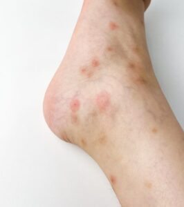
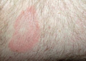
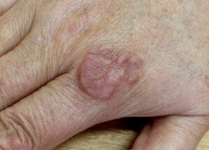
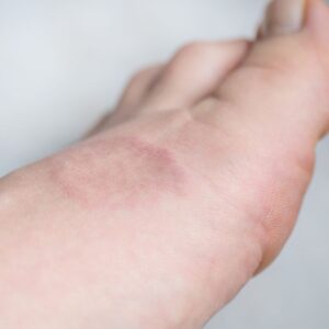
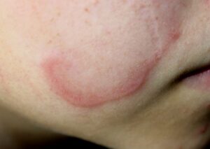
Localized GA (Most Common Form) | Presents as isolated, ring-shaped or circular patches with a raised, smooth, and firm border surrounding a completely flat, clear center [cite: 1074, 1075, 1079]. The border is composed of tiny individual skin-colored, pink, or light purple (violaceous) bumps arranged in a neat ring [cite: 1075, 1155]. This variant typically resolves on its own without leaving any scars [cite: 1109]. |
| Generalized (Disseminated) GA | Marked by widespread, numerous, and scattered individual small bumps or plaques populating broad areas of the body simultaneously [cite: 1081, 1132]. This subtype is statistically much more persistent, can last for multiple decades, and is more frequently encountered in older adult populations [cite: 1086, 1119]. |
| Subcutaneous GA | An asymptomatic variant seen almost exclusively in young pediatric patients [cite: 1080]. Instead of a surface rash, it forms deep, firm, non-tender nodules hidden completely underneath the skin layer, typically over bony regions like the lower legs or scalp [cite: 1080, 1143, 1150]. |
| Perforating GA | A rare, specialized variant where the degenerating dermal collagen fibers are actively pushed upward and eliminated directly through the outer skin layer (epidermis) [cite: 1106, 1133]. This process forms small bumps featuring a dimpled, pitted, or crusted central core [cite: 1107, 1154]. |
Am I at risk for Granuloma Annulare or underlying conditions?
Experiencing a breakout of granuloma annulare is an isolated skin phenomenon [cite: 1074]. It has absolutely zero connection to personal hygiene flaws, skin cleanliness, or a globally deficient immune system.
- The Age and Gender Axis: GA demonstrates a clear statistical preference for children, teenagers, and young adults under 30 years of age (except for the generalized form, which skews older) [cite: 1086]. It exhibits a clear 2:1 female-to-male demographic predominance [cite: 1087].
- Systemic Health Connections: While the skin lesions themselves are entirely harmless, medical literature has established clear statistical associations between GA and several internal systemic conditions [cite: 1084]. These include **diabetes mellitus, autoimmune thyroiditis, and hyperlipidemia** (elevated cholesterol/triglycerides) [cite: 1084, 1137]. Screening for these conditions can be an important component of your broader care plan [cite: 1084].
- Targeted Ancestral Risks: Specific demographic variants exist; individuals of **Ethnic Hawaiian** descent are documented to face a statistically higher clinical risk for developing the rare, crusting perforating variant of GA [cite: 1088].
- Predictable Recurrence Rates: Even after the lesions disappear completely, your skin barrier lines maintain a **40% recurrence rate** [cite: 1117]. Interestingly, these recurrent flares show a clear biological tendency to return to the original sites, but they clear significantly faster than the primary outbreak [cite: 1118].
Where and How It Appears on My Body
Granuloma Annulare populates highly predictable landmarks across your skin surface while preserving a smooth, scale-free profile that sets it apart from fungal or scaling rashes [cite: 1074, 1096].
- The Extremity Anchor: GA shows a massive preference for the extremities [cite: 1078]. Localized forms focus heavily on the fingers, the backs of the hands, the tops of the feet, the ankles, and directly over one or both elbows [cite: 1079]. Generalized variants will actively expand to involve the trunk and neck lines [cite: 1081].
- The Smooth, Scale-Free Border: Unlike ringworm (tinea), a classic GA plaque is completely smooth to the touch and **lacks any surface peeling, flaking, or fine scale** [cite: 1074, 1096]. The outermost ring sits slightly elevated, forming a firm, circular rim that is visibly darker or more violaceous than the clear, flat center [cite: 1075, 1155].
- Muted Complexion Clues: On deeper skin complexions, the classic pink or violaceous hue can appear muted, dusky brown, or entirely flesh-colored, making the ring border harder to spot in dim lighting [cite: 1075, 1076]. On skin of color, it is crucial to use proper side-lighting (or a magnifying device) to trace the raised ring pattern accurately [cite: 1075, 1090].
Solutions I Can Try at Home
Because granuloma annulare is a self-contained, internally driven dermal reaction pattern, home care is focused entirely on preventing unnecessary tissue stress and managing localized symptoms safely [cite: 1083, 1109].
- Practice Triage and Patience: This is the single most vital tool [cite: 1109]. Up to 50% of all cases of granuloma annulare undergo complete, **spontaneous resolution within 2 years** without a single medical intervention [cite: 1117]. If the spots do not itch or cause social distress, simply leaving them alone to heal naturally is a highly valid approach [cite: 1109, 1123].
- Do Not Scrub or Exfoliate the Rings: Do not use harsh loofahs, abrasive body scrubs, or chemical exfoliating acids on active rings. Because GA involves a deep breakdown of dermal collagen rather than a surface cell buildup, scrubbing will only irritate the tissue lines and cause the ring to inflame further [cite: 1083, 1128].
- Keep Skin Well-Lubricated: Apply thick, fragrance-free emollient creams to the areas daily. Maintaining a well-hydrated outer layer (epidermis) reduces any superficial skin tension and keeps the skin barrier resilient against external friction [cite: 1133].
When Should I See a Dermatology Provider?
GA can easily mask itself as common ringworm, lichen planus, or an insect bite [cite: 1095, 1096, 1099]. Seeking professional clinical triage ensures a precise diagnosis through an office **4mm punch skin biopsy** to evaluate your tissue layers under a microscope and rule out dangerous lookalikes [cite: 1127].
Seek Professional Help if You Notice These Warning Signs:
- Diagnostic Uncertainty or Atypical Expansion: Your rash features an irregular, ill-defined shape or lacks a clear circular border, requiring a skin biopsy to definitively distinguish it from the arcuate plaques of mycosis fungoides (a type of cutaneous lymphoma) [cite: 1097].
- Widespread, Persistent Eruptions: The smooth bumps multiply rapidly across your trunk, arms, and neck (generalized GA), causing severe cosmetic distress or persisting past several years without signs of flattening [cite: 1081, 1114, 1119].
- The Deep, Rapidly Growing Nodule: You or your child notice a hard, fixed, lump growing deep underneath the skin surface or on a joint line, requiring professional correlation to rule out a rheumatoid nodule or an epithelioid sarcoma [cite: 1105, 1143, 1150].
Frequently Asked Questions
- Q: What primary medical options will my provider use to clear localized patches of GA?
A: If your skin lesions are persistent, itchy, or cosmetically obtrusive, your provider will initiate targeted localized therapies [cite: 1110]. First-line clinical interventions include prescription-strength **topical corticosteroid creams** or non-steroidal steroid-sparing agents applied directly to the active borders [cite: 1111, 1129]. To accelerate clearance for stubborn, thickened patches, your dermatologist can perform in-office **intralesional corticosteroid injections** (such as triamcinolone acetonide) directly into the raised ring to rapidly calm the local inflammatory cells and halt collagen degeneration [cite: 1112, 1139]. Localized destruction methods like cryotherapy (freezing) or laser ablation can also be considered [cite: 1113, 1130]. - Q: What advanced systemic medications are available if my GA is widespread or generalized?
A: For severe, disseminated, or generalized variants that cover a high percentage of your body surface, localized creams are impractical, requiring your provider to step up to systemic therapies [cite: 1114, 1132, 1151]. Advanced systemic options include oral medications such as **methotrexate, cyclosporine, dapsone, hydroxychloroquine, or oral retinoids** (isotretinoin) [cite: 1115]. Modern targeted biological therapies, specifically **TNF-alpha inhibitors** (which block an essential inflammatory protein driving the disease), have also demonstrated high efficacy rates in clearing recalcitrant cases [cite: 1115, 1152]. - Q: How do pathologists distinguish GA from common fungal ringworm under a microscope?
A: Under a microscope, your pathologist can easily execute a definitive separation because the two conditions possess entirely distinct tissue signatures [cite: 1093]. Fungal ringworm (tinea) is a superficial infection confined entirely to the outer skin layer, whereas granuloma annulare leaves a deep signature in the dermis [cite: 1096, 1131]. A skin biopsy of GA explicitly reveals **necrobiotic degeneration of dermal collagen** surrounded by a neat palisade of histiocytes and helper T-lymphocytes [cite: 1091, 1128]. This specialized microscopic landscape is completely absent in tinea, ensuring total diagnostic certainty [cite: 1093, 1096].
The long-term outlook for the vast majority of individuals with localized granuloma annulare is outstanding, with half of all cases clearing completely and beautifully on their own within two years [cite: 1117]. Success relies on patience during the natural healing cycle, strict adherence to your prescribed localized or systemic routine, and a close, collaborative partnership with your interprofessional dermatology team to keep your body healthy and fully protected [cite: 1109, 1114, 1123].