Erythema Multiforme (EM) is an acute, immune-mediated skin condition characterized by the sudden appearance of distinctive, ring-shaped spots known as "target" lesions [cite: 1882, 1944]. While the abrupt arrival of a widespread, multi-colored rash can look alarming, EM is typically a self-limiting reaction that resolves naturally without leaving permanent scars [cite: 1935, 1941]. Understanding what triggers this immune response is the first strategic step toward securing targeted symptom relief, managing recurrences, and protecting your long-term skin health [cite: 1937, 1938, 1948].
What Is Causing This Sudden, Target-Shaped Rash?
Understanding how your body’s immune pathways react to internal factors is a strategic first step in managing erythema multiforme. EM is fundamentally an immune-mediated disorder, meaning it represents an overactive defense response launched by your cells [cite: 1893, 1944]. It does not arise from poor personal hygiene or a lack of personal cleanliness.

Instead, the inflammatory cascade is kicked off by a reaction to an external or internal antigen [cite: 1893]. The vast majority of cases are triggered by common infectious agents rather than food allergies or surface chemicals [cite: 1893]. The primary drivers include:
- Viral Infections: The Herpes Simplex Virus (HSV) accounts for the absolute majority of cases [cite: 1894]. It is important to note that the virus can trigger EM even if your cold sores are completely subclinical or unnoticeable [cite: 1896]. Other viral triggers include Cytomegalovirus, Epstein-Barr Virus, and the Influenza virus [cite: 1898].
- Bacterial Microbes: Mycoplasma pneumoniae serves as a major infectious trigger, particularly in children and adolescents [cite: 1897].
- Medication Exposures: Certain prescription drugs can prompt an EM reaction, including specific antibiotics, anti-epileptic (seizure) medications, and non-steroidal anti-inflammatory drugs (NSAIDs) [cite: 1899, 1900].
You can think of an EM eruption as an internal “immune alarm” that manifests visually on the surface of your skin [cite: 1944]. Identifying this condition early through an expert physical examination is the essential “So What?” factor in your recovery plan [cite: 1912]. Pinpointing the condition prevents medical confusion and ensures that dangerous, severe lookalikes are safely ruled out from the beginning [cite: 1933, 1947].
Understanding the Visual Timeline: Tracking the Classic Target Lesion
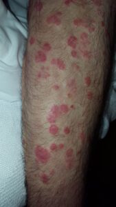
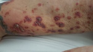
Erythema multiforme features a highly specific, three-zone physical pattern that develops with striking speed [cite: 1884, 1886]. Recognizing this hallmark pattern helps you monitor your condition accurately [cite: 1913].
| Lesion Zone | Visual Appearance and Architectural Features |
|---|---|
The Central Core (Inner Zone) | A dark, dusky area indicating localized epidermal tissue death (necrosis) [cite: 1884]. In severe presentations, this central core can split open into a fluid-filled blister (bulla) or develop a hard crusted scab [cite: 1885]. |
The Intermediate Ring (Middle Zone) | A pale, lighter-colored, and slightly swollen (edematous) ring that circles the dark core, creating a clear contrast zone [cite: 1884]. |
The Outer Margin (Peripheral Zone) | A bright red, flat or slightly raised inflammatory border (erythematous margin) that outlines the entire spot, completing the classic target shape [cite: 1883, 1884]. |
Am I at risk for erythema multiforme or recurrences?
Developing erythema multiforme is an immune-mediated event [cite: 1944]. It skews toward specific age groups and underlying medical histories [cite: 1906, 1908].
- The Young Adult Profile: While it can technically surface at any age, EM demonstrates a clear clinical preference for young adults between the ages of 20 and 40, showing a very slight predominance in males over females [cite: 1906, 1907]. It is uncommon in early childhood [cite: 1906].
- The Recurrence Path: If you have experienced an episode of EM in the past, you face a statistically higher risk for developing recurrent outbreaks, especially if your initial eruption was triggered by an underlying Herpes Simplex Virus infection [cite: 1908, 1942].
- Associated Health Flags: Susceptibility lines are mildly influenced by systemic conditions, such as underlying autoimmune disorders, inflammatory bowel disease (IBD), Hepatitis C, or a state of systemic immunosuppression [cite: 1902, 1909].
- Sparing Status Clues: Remarkably, a classic case of uncomplicated erythema multiforme remains a purely cutaneous and localized event; it is **not accompanied by an internal fever, swollen lymph nodes (lymphadenopathy), or liver enlargement (hepatosplenomegaly)** [cite: 1887]. Finding these systemic features means your rash has a completely different cause [cite: 1925, 1928].
Where and How It Appears on My Body
EM lesions follow a highly symmetric anatomical map across your limbs, presenting unique landmarks that help your provider instantly separate it from standard hives or viral measles [cite: 1889, 1913, 1921, 1925].
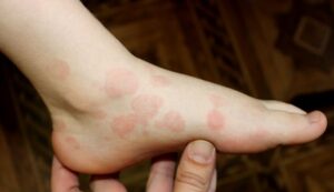
- The Distant Extensor Anchor: The rash shows an overwhelming biological preference for the extensor surfaces of the distal upper extremities [cite: 1889]. Lesions cluster heavily on the backs of the hands (dorsal hands) and the forearms [cite: 1890]. Secondary sites include the palms, neck, face, and trunk [cite: 1890].
- The Fixed 7-Day Rule: Unlike common hives (urticaria) which move dynamically and vanish from a spot within 24 hours, individual EM target spots are completely **fixed and stay anchored in the exact same location for at least 7 days** [cite: 1921].
- The Blaschko Eruptive Burst: The onset of the rash is incredibly abrupt [cite: 1886]. The majority of your spots (which usually number over 100 lesions across the body) explode onto the skin within the first 24 hours, and virtually all patches arrive within a strict 72-hour window [cite: 1886, 1887].
- Muted Complexion Clues: On deeper skin complexions, the bright red outer ring can look muted, dusky purple, or darkly hyperpigmented [cite: 1884, 1968]. In skin of color, tracing the sharp, three-zone circular border remains the primary clinical anchor used to locate active spots [cite: 1884, 1913].
Solutions I Can Try at Home
Because erythema multiforme is an internally driven immune response, basic skin lotions provide limited relief [cite: 1893]. At-home support focuses on gentle care and tracking your symptoms safely [cite: 1912, 1938].
- Practice Triage and Patience: This is a highly encouraging tool [cite: 1935]. Uncomplicated EM is a classic self-limiting event that is pre-programmed to undergo complete, natural resolution within a **4 to 6 week window** [cite: 1935].
- Gently Log Your Symptoms: Keep a careful written record of your breakout [cite: 1912]. Note exactly how quickly your spots appeared and document whether you had a recent cold sore or a dry, hacking respiratory cough in the weeks preceding the rash, which provides your clinical team with essential diagnostic triggers [cite: 1895, 1897].
- Do Not Manually Pop Central Blisters: If the central core of your target spots develops a fluid-filled blister, leave it completely alone [cite: 1885]. Manually popping or squeezing the blister breaks your surrounding skin barrier lines, introduces surface bacteria, and delays your skin’s healing path [cite: 1970].
When Should I See a Dermatology Provider?
Erythema multiforme can easily mask itself as a fixed drug eruption or a serious bacterial infection [cite: 1929, 1930]. Seeking professional clinical triage early ensures you secure an accurate diagnosis and rules out dangerous, life-threatening lookalikes [cite: 1933, 1947].
Seek Professional Help immediately if You Notice These Warning Signs:
- Diagnostic Uncertainty or Atypical Shapes: Your rash lacks clear three-zone target spots or has irregular, dusky borders, requiring an expert **4mm punch skin biopsy** to evaluate your tissue layers under a microscope and establish absolute clarity [cite: 1916, 1926].
- The Emergence of Painful Mouth Sores: Active skin lesions are accompanied by significant, painful sores or raw erosions inside your mouth, lips, or throat (mucous membrane involvement) [cite: 1891, 1927].
- Prominent Systemic Illness: Your skin eruptions are accompanied by a high fever, severe malaise, or a raw sloughing and peeling of your skin, indicating a potential progression toward **Stevens-Johnson Syndrome (SJS) or Toxic Epidermal Necrolysis (TEN)**—which are severe medical emergencies requiring immediate hospitalization [cite: 1926, 1927, 1928, 1933, 1939].
Frequently Asked Questions
- Q: What extensive laboratory panel is executed if my diagnosis is unclear?
A: In unclear or atypical cases, your provider will order a thorough blood panel to evaluate your systemic health and isolate potential infectious triggers [cite: 1912, 1917]. This workup includes a Complete Blood Count (CBC), a Comprehensive Metabolic Panel (CMP-14), and an Antinuclear Antibody (ANA) screening [cite: 1918]. To hunt for the specific root cause, your team will order **Herpes Simplex Virus (HSV) PCR or serology**, alongside **Mycoplasma pneumoniae IgM serology or PCR testing** [cite: 1914, 1915]. Your panel will also track inflammatory markers (ESR/CRP) and check your serum **complement levels**, which are uniquely normal in EM but abnormally low in lookalike conditions like urticarial vasculitis [cite: 1918, 1932]. - Q: What primary medical treatments are used to manage widespread itching and discomfort?
A: For an active, uncomplicated outbreak, medical management focuses entirely on symptom reduction and skin soothing [cite: 1938]. Your provider will prescribe **oral antihistamines** to calm systemic itch pathways [cite: 1938]. This is routinely paired with a large, **454-gram jar of Triamcinolone acetonide 0.1% topical corticosteroid cream** [cite: 1938]. This large volume allows for safe, widespread application to the extensor surfaces of your limbs twice daily to rapidly cool localized inflammation and ease skin discomfort [cite: 1889, 1938]. - Q: What specialized treatment algorithm exists if I suffer from chronic, recurrent EM?
A: If you suffer from frequent, disruptive recurrences driven by the Herpes Simplex Virus, your dermatology team will initiate a long-term preventative routine [cite: 1937, 1942]. Rather than waiting for a rash to erupt, your provider will place you on continuous, **prophylactic antiviral therapy (such as oral acyclovir or valacyclovir) maintained strictly for 6 to 12 months** [cite: 1937]. This continuous suppressive approach safely blocks subclinical viral replication, effectively breaking the inflammatory highway and preventing future target rash flares [cite: 1896, 1937].
The long-term outlook for the vast majority of individuals with erythema multiforme is outstanding, with cutaneous lesions clearing completely and beautifully on their own within four weeks without any permanent scarring [cite: 1935, 1941]. Success relies on identifying your specific infectious trigger, strict adherence to your localized soothing or antiviral routine, and maintaining a close, collaborative relationship with your interprofessional dermatology team to keep your body fully protected [cite: 1936, 1937, 1948].