Quick Summary: Lymphomatoid papulosis (LyP) is a rare, chronic skin disorder characterized by recurrent crops of self-healing bumps that can develop scabs or open sores. Although it is classified as a low-grade lymphoproliferative disorder, it carries an excellent long-term survival rate, meaning primary management focuses on suppressing active skin breakouts, managing symptoms, and maintaining regular routine monitoring.
What Is Causing These Self-Healing Bumps?
Understanding how your body’s immune surveillance network operates within the skin is a strategic first step in managing your condition. Lymphomatoid papulosis is classified as a rare, self-healing primary cutaneous CD30+ T-cell lymphoproliferative disorder [cite: 3302, 3335]. This means it involves an abnormal, localized increase of specific immune cells—specifically CD30+ T-lymphocytes—directly within the skin layers [cite: 3303, 3343, 3344].

The root cause or exact pathophysiology driving LyP is largely unknown, though research suggests genetic factors may play a role [cite: 3301, 3304, 3347]. You can think of an eruptive crop as a temporary “cellular traffic jam” where these overactive white blood cells cluster in the skin, cause a localized tissue breakdown, and then naturally disperse [cite: 3293, 3295]. Identifying this condition early through a professional clinical assessment is the essential “So What?” factor in your care plan [cite: 3309]. Because it is an internally driven immune-mediated process rather than a simple surface allergy, establishing a precise diagnosis prevents medical guesswork and ensures your skin receives pathway-specific, suppressive management [cite: 3319, 3339].
Understanding the Visual Timeline: The Life Cycle of an LyP Nodule
Lymphomatoid papulosis has a highly unique biological hallmark: individual spots are pre-programmed to auto-involute and completely disappear on their own over several weeks [cite: 3295, 3380]. Recognizing this waxing and waning baseline timeline helps you monitor changes safely [cite: 3297].
| Lesion Stage | Key Characteristics and Physical Layouts |
|---|---|
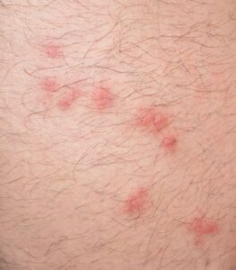
| Active Eruption | Presents as a sudden crop of small, firm, reddish-brown bumps (papules) [cite: 3293, 3342]. Over days, they often develop central cell death (necrosis), dark bloody scabs (crusting), or break down into shallow, open ulcers [cite: 3294, 3345]. Bumps can occasionally be mildly itchy or tender [cite: 3317, 3376]. |
| Spontaneous Regression | Without any therapeutic intervention, the individual crusted nodule flattens out, stops scaling, and spontaneously resolves completely within a strict 2 to 8 week window [cite: 3295]. |
| The Healing Mark | As the active sore clears, it frequently leaves behind temporary light or dark shadows (post-inflammatory pigmentary changes) or tiny pockmarked depressions known as atrophic scarring [cite: 3296, 3358, 3359]. |
Am I at risk for lymphomatoid papulosis or internal lymphoma?
Developing lymphomatoid papulosis is an isolated, random biological event [cite: 3288]. It has absolutely zero connection to personal hygiene flaws, a lack of cleanliness, or dietary mistakes.
- The Age Profile: LyP can develop at any age, but it exhibits a strong statistical preference for adults, with the peak clinical incidence occurring during your fifties (the fifth decade of life) [cite: 3306]. It is rare in young pediatric populations [cite: 3306].
- The Chronic Waxing and Waning Course: LyP typically follows a chronic, persistent course that can wave and recur over several months, years, or even multiple decades [cite: 3331]. When treatments are started and later stopped, the papules routinely return [cite: 3322].
- Associated Lymphoma Risks: It is clinically essential to understand that up to 20% of patients diagnosed with LyP may develop an associated secondary cutaneous or systemic lymphoma over their lifetime [cite: 3307, 3332]. The most common associated lymphomas include mycosis fungoides (MF), primary cutaneous anaplastic large cell lymphoma (PC-ALCL), or Hodgkin lymphoma [cite: 3332].
- Favorable Survival Baseline: Despite this associated risk, the overall long-term prognosis for LyP is exceptionally outstanding and favorable [cite: 3332, 3340]. Large-scale clinical studies demonstrate an incredibly high survival rate, with a very low percentage of cases directly leading to serious systemic complications [cite: 3333].
Where and How It Appears on My Body
LyP leaves a highly scattered but characteristic physical layout across the skin lines that helps your healthcare provider differentiate it from common external conditions.
- Trunk and Extremity Prevalence: The crops of papules demonstrate a massive preference for the skin of the chest, abdomen, back, and the limbs [cite: 3299].
- Multi-Stage Mixed Presentation: Because new crops burst forth while older ones are shrinking, you will frequently see fresh red bumps resting immediately adjacent to dark crusted ulcers and old atrophic scars, creating a highly mottled visual profile [cite: 3294, 3295, 3296].
- Muted Color Clues: On deeper skin complexions, the classic reddish-brown hue of the active spots may look muted, deep purple, or dark brown [cite: 3377]. In skin of color, the temporary light marks left behind by regressing spots can be highly visible against your baseline skin tone [cite: 3358].
Solutions I Can Try at Home
Because lymphomatoid papulosis is driven by an internal, lymphoproliferative process, home management focuses entirely on protecting your skin lines and tracking active lesions carefully [cite: 3302, 3320].
- Enforce the 8-Week Spot Test: If a new reddish-brown bump arrives, note its date of arrival and lightly track its location. Document whether that specific enclosed nodule flattens, auto-involutes, and clears on its own within 8 weeks [cite: 3295]. Confirming this spontaneous self-healing cycle is a highly valuable clinical clue for your medical team [cite: 3311, 3337].
- Do Not Pick Crusted Scabs: Avoid aggressively picking or scratching away the necrotic central scabs or open sores [cite: 3294]. Picking will not stop the underlying T-cell clustering; it will only break your skin barrier lines, prolong the scarring path, and risk introducing a secondary bacterial infection [cite: 3303, 3359].
- Maintain a Clear Photographic Diary: Take clear, well-lit photos of your trunk and limbs monthly. This objective record helps your interprofessional team distinguish whether your spots are successfully resolving or behaving abnormally [cite: 3328, 3348].
When Should I See a Dermatology Provider?
Lymphomatoid papulosis cannot be diagnosed by sight alone. Because early lesions can look identical to common insect bites or severe eczema, your provider must perform a safe, in-office 4mm punch skin biopsy to evaluate the tissue layers under a microscope and establish absolute diagnostic clarity [cite: 3309, 3317].
Seek Professional Help if You Notice These Warning Signs:
- The Non-Regressing Nodule: A single, isolated LyP nodule continues to steadily enlarge, grows larger than a standard spot, and completely fails to spontaneously flatten or resolve after 8 weeks [cite: 3329]. This is a critical clinical warning indicator that requires an immediate repeat punch biopsy to rule out the development of an aggressive anaplastic CD30+ large cell lymphoma [cite: 3329].
- Diagnostic Uncertainty: You are experiencing a chronic, recurring crop of crusted or necrotic skin bumps that have completely failed to improve with standard over-the-counter moisturizers or basic anti-itch creams [cite: 3293, 3294, 3320].
- The Emergence of Widespread Plaquing: Your skin changes begin forming extensive, persistent flat patches or thickened plaques that do not clear up on their own, requiring careful histological review to evaluate for an overlapping mycosis fungoides process [cite: 3316, 3374].
- Constitic Organ Signaling: Your skin eruptions are suddenly accompanied by systemic constitutional symptoms, including recurrent unexplained fevers, sudden night sweats, unprompted weight loss, or firm, swollen lymph nodes in your neck, armpits, or groin creases [cite: 3328, 3368].
Frequently Asked Questions
- Q: How do providers histologically separate LyP from other severe conditions under a microscope?
A: Under a microscope, your pathologist will utilize highly advanced immunohistochemistry (IHC) staining to analyze your cells [cite: 3348, 3366]. A diagnostic hallmark of lymphomatoid papulosis is the clear presence of atypical T-cells that stain strongly positive for the CD30 protein receptor [cite: 3303, 3343, 3352]. These specific cells are completely absent or minimal in benign lookalikes like standard pityriasis lichenoides (PL) or simple arthropod insect bites, allowing for definitive clinicopathological separation [cite: 3314, 3317, 3348]. - Q: What primary suppressive medical treatments are prescribed if my disease is extensive or scarring?
A: For mild, limited disease that does not cause physical scarring or distress, medical treatment is completely optional and simple observation may be preferred [cite: 3321]. However, if treatment is initiated to suppress extensive outbreaks, first-line options include the application of superpotent Class I topical steroid ointments or specialized 1% bexarotene gels directly to early, active papules [cite: 3323, 3324]. For widespread or cosmetically obtrusive cases, highly effective low-dose weekly oral Methotrexate (5 to 10 mg per week) or structured in-office narrowband UVB or PUVA phototherapy sessions can be utilized to successfully suppress the active crops [cite: 3325, 3326]. - Q: Can I permanently cure LyP, and what does long-term wellness tracking look like?
A: Currently, there is no permanent cure for lymphomatoid papulosis, and the primary focus of modern care is symptom suppression and lesion control [cite: 3320]. Because of your statistically elevated lifetime risk for secondary cutaneous or systemic malignancies, maintaining a lifetime, regular follow-up surveillance schedule with your interprofessional dermatology team is absolutely vital to protect your long-term health [cite: 3307, 3328]. Routine laboratory blood tests or internal imaging markers are typically completely normal and are not required unless your physical clinical checkup highlights systemic warning signals [cite: 3312].
The long-term outlook for healthy individuals with lymphomatoid papulosis remains incredibly favorable, with an outstanding survival profile and excellent general health outcomes [cite: 3332, 3333]. Success relies on early clinicopathological verification via an office biopsy, strict behavioral tracking of lesion life cycles, and establishing a regular, ongoing relationship with your dedicated dermatology care team to keep your body protected [cite: 3309, 3328, 3340].