Quick Summary: An infantile hemangioma (strawberry nevus) is a common, benign vascular tumor of infancy characterized by a dense grouping of extra blood vessels. While the vast majority naturally shrink and disappear completely on their own, early professional clinical mapping is essential to identify high-risk hotspots that require low-risk, safe medical treatments.
What Is Causing This Red Birthmark?
Understanding the unique, time-limited lifecycle of your infant’s skin cells is a strategic first step in managing an infantile hemangioma. This condition occurs when an abnormally dense group of extra blood vessels multiplies rapidly in a localized area. You can think of this birthmark as a temporary “vascular milestone” rather than a dangerous or invasive tumor; current research suggests its development may be linked to specific proteins produced by the placenta during pregnancy.


The root cause is an internal, biological proliferation that typically begins within the first few weeks of life. Tracking this birthmark early is the “So What?” factor in your child’s care, allowing you to establish whether the hemangioma is a low-risk spot that can be safely observed or a high-risk variant that qualifies for non-surgical, protective medical therapy.
Understanding the Phases: Proliferation vs. Involution
Infantile hemangiomas follow a highly structured lifecycle, moving from rapid growth to complete natural fading. Recognizing these phases prevents unnecessary parenting anxiety.
| Lifecycle Phase | Timeline and Structural Characteristics |
|---|---|
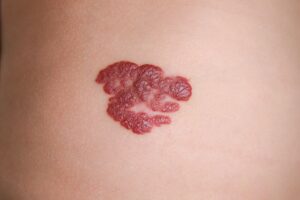
| Proliferative (Growth) | Timeline: First year of life. Appearance: The red mark grows rapidly, transforming into a raised, spongy mass that can protrude prominently from the skin surface. |
| Involution (Shrinking) | Timeline: Fades predictably with age. 50% clear by age 5, 70% by age 7, and nearly 100% resolve by age 10. Appearance: The bright color fades completely, leaving normal skin or faint discoloration. |
Am I at risk for complications?
Developing a solitary infantile hemangioma is incredibly common and has absolutely zero connection to pregnancy choices, maternal diet, or infant hygiene. Susceptibility depends heavily on the structural type and anatomical layout of the growth.
- Special Site Hotspots: Hemangiomas involving the tip of the nose, the lip, or the ear area carry an increased risk of slow natural shrinkage and permanent textural scarring, making early intervention a clinical priority.
- Segmental Formations: Large hemangiomas that cover a broad, continuous segment of the face, neck, or lower back carry higher complication rates due to the potential for associated internal anomalies.
- Friction Points: Growths situated in skin folds or areas prone to mechanical rubbing (like the diaper area or limbs) are at high risk for surface breakdown or ulceration.
Where and How It Appears on My Child’s Body
Infantile hemangiomas populate specific zones on the body, presenting with visual landmarks that change based on depth.
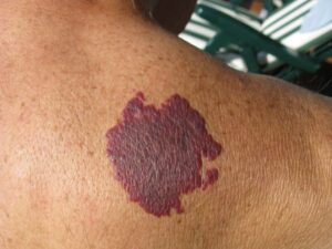
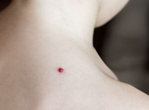
- Head and Neck Prevalance: The face, scalp, and back of the neck represent the most common hotspots, often presenting as a single, isolated, bright red “strawberry” plaque.
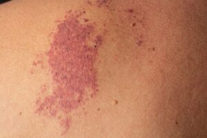
- The Deep Profile: Deeper hemangiomas can present beneath the surface as a soft, violaceous or bluish bulge, sometimes showing an underlying patch of tiny broken blood vessels surrounded by a pale ring.
- The “Airway Warning” Layout: Hemangiomas located in a “beard distribution” (jawline, chin, lower lip, and front of the neck) carry a unique structural risk, as they can be associated with subglottic hemangiomas that compromise breathing between 6 and 12 weeks of life.
Solutions I Can Try at Home
Because the vast majority of hemangiomas complete their lifecycle beautifully without intervention, home care focuses on strict protective observation and skin barrier support.
- Regular Photo-Documentation: Take clear, well-lit pictures of the birthmark at the same time each month alongside a small ruler. This provides your medical team with the precise objective evidence needed to monitor its growth trajectory.
- Do Not Squeeze or Compress: Never attempt to squeeze, pop, or use tight homemade compression bands on the spongy mass, as this can injure the fragile blood vessels and cause severe ulceration.
- Protect from Water and Friction: Keep skin folds dry, use mild suds-free cleansers, and apply a lightweight protective ointment around high-friction spots to shield the delicate tissue barrier.
When Should I See a Dermatology Provider?
While patience is the rule, professional clinical triage must be initiated immediately if a hemangioma develops warning signs or tracks into a cosmetically or functionally sensitive zone.
Seek Professional Help immediately if You Notice These “Red Flags”:
- The “Early White” Warning: You notice a faint, whitish discoloration developing across the surface of a bright red hemangioma. This is a critical clinical indicator that the lesion is about to ulcerate, which can cause intense pain, bleeding, and deep scarring if not managed clinically.
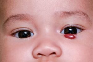
- Functional Impediment: The growth is located near the eye (periorbital area), which can cause visual deficits like astigmatism or lazy eye (amblyopia); or it involves the lip, hands, or knees, interfering with normal feeding or crawling.
- Respiratory Signs: A child with a beard-distribution hemangioma develops a persistent cough, hoarseness, stridor (high-pitched breathing), or respiratory distress.
- Large Facial Segments: A broad, segmental hemangioma involves the face, requiring a formal workup (including ophthalmology, echocardiogram, and head MRI) to rule out PHACE syndrome before initiating oral therapy.
Frequently Asked Questions
- Q: How does oral Propranolol work to clear high-risk hemangiomas?
A: For high-risk or functionally sensitive lesions, oral Propranolol is the preferred, safe, non-surgical approach. It works directly by constricting the extra blood vessels and reducing blood flow to shrink the tumor safely during its growth period. It is typically dosed twice daily with food up to 13 months of age, using a careful two-week taper when stopping treatment. - Q: What happens if a child on Propranolol catches a cold?
A: If your child develops an upper respiratory infection (URI) with wheezing or coughing, you should stop the Propranolol immediately, as beta-blockers can increase breathing difficulties. Once the infection has fully resolved, you can safely restart the treatment under your provider’s guidance. - Q: When are advanced therapies like lasers or surgery considered?
A: Pulsed dye lasers (PDL) can be used effectively to treat painful, ulcerated lesions or to clear residual surface blood vessels (telangiectases) after the growth phase is complete. Surgical excision is reserved for complex scenarios, such as ulcerated scalp lesions or spots with a very high risk of permanent facial disfigurement.
The long-term outlook for infantile hemangiomas is outstanding, with over half of all children demonstrating completely normal, unmarked skin by their tenth birthday. Success relies on consistent monthly photo-tracking, protecting the fragile skin barrier, and partnering with your clinical team to initiate targeted therapies early if a high-risk zone is involved.