Quick Summary: Dermatomyositis (DM) is a rare, complex autoimmune disease characterized by unique skin rashes and symmetrical muscle weakness [cite: 2024, 2101]. While it frequently impacts both the skin and the muscles, some individuals experience the classic skin changes without ever developing active muscle disease [cite: 2041, 2101]. Early clinical diagnosis supported by a professional skin biopsy is vital to control active inflammation, initiate screening for underlying internal conditions, and safely protect your long-term health [cite: 2065, 2093, 2096, 2102].
What Is Causing My Sensitive Skin Rashes and Muscle Weakness?
Podcast:
Learn More About Dermatomyositis
Instead, the inflammatory cascade is believed to be kicked off by a complex combination of external factors—such as an underlying malignancy, specific medications, or infectious agents—acting in individuals who carry a distinct genetic predisposition [cite: 2061]. When this internal highway is activated, it triggers specialized inflammatory pathways that damage blood vessels and connective tissues in your skin and muscles. You can think of an active DM flare-up as an internal “system-wide cellular misfire.” Identifying this condition early through an expert clinical evaluation is the essential “So What?” factor in your care plan [cite: 2102]. Because it operates via deep systemic autoimmune networks, available skin therapies are frequently inadequate on their own, and standard over-the-counter soothing lotions cannot stop the cycle, making path-specific medical treatments and regular multidisciplinary tracking mandatory to defend your tissue lines [cite: 2087, 2102].
Understanding the Condition: Comparing Dermatomyositis to Systemic Lupus
Because dermatomyositis causes prominent facial flushing and sun-sensitive rashes, it is frequently confused with another systemic autoimmune condition called Systemic Lupus Erythematosus (SLE) [cite: 2074]. Recognizing these key clinical differences helps your healthcare provider establish the correct diagnostic path early [cite: 2029, 2074].
| Autoimmune Condition | Key Differentiators, Anatomy, and Visual Configurations |
|---|---|
| Systemic Lupus (SLE) | Facial Layout: Features a classic “butterfly” rash that covers the cheeks and the bridge of the nose but strictly spares the nasolabial folds (the creases running from the nose to the corners of the mouth) [cite: 2029, 2074]. Features & Hands: If the hands are involved, the rash characteristically spares the knuckles [cite: 1791]. Rashes typically display a red poikiloderma and are significantly less itchy (not integral to the disease) [cite: 2027, 2074]. |
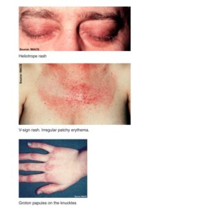
| Dermatomyositis (DM) | Facial Layout: Facial redness actively involves the nasolabial folds and the eyelids, often presenting as a distinctive violet or lilac discoloration called a heliotrope rash [cite: 2029, 2032, 2113]. Features & Hands: Displays a distinct violet poikiloderma (a mixture of dark and light spots, visible spider veins, and thinned skin) [cite: 2027, 2116]. The skin changes, knuckles, and the scalp are often intensely itchy (pruritic), which helps distinguish it from lupus [cite: 2033, 2035, 2074]. |
Am I at risk for internal organ complications or cancer?
Developing dermatomyositis is an internal autoimmune event [cite: 2060]. It affects individuals across all age ranges, but your specific age baseline changes your expected clinical profile and long-term risk tracking [cite: 2026, 2043].
- The Gender Predominance: DM is a relatively rare condition that demonstrates a clear demographic preference, affecting women two to three times more frequently than men [cite: 2063].
- The Juvenile vs. Adult Split: The disease presents in two primary forms [cite: 2026]:
- Juvenile Form: Tends to cause more muscle inflammation but is not associated with internal cancer [cite: 2044]. However, children face a high risk of developing calcinosis cutis, affecting between 25% and 70% of patients, which forms hard, painful calcium nodules under the skin that can drain a chalky material [cite: 2037, 2038, 2040, 2108].
- Adult Form: Carries a powerful statistical association with hidden internal cancers (malignancies) and progressive lung disorders [cite: 2045, 2103].
- The Occult Malignancy Risk: Because adult-onset DM can act as a secondary reaction to a hidden tumor, adults face a high risk of an underlying cancer [cite: 2061]. This requires rigorous cancer screenings at the time of diagnosis and repeat checks every 3 years if initial tests are negative [cite: 2093].
- The Cardiac and Pulmonary Threat: Pulmonary disease occurs in approximately 15% to 30% of adult patients, generally presenting as a diffuse interstitial fibrosis [cite: 2050]. Crucially, hidden cardiac involvement represents a major negative prognostic factor for survival, making thorough evaluation essential to protect your health [cite: 2051, 2098, 2104].
Where and How It Appears on My Body
Dermatomyositis leaves an unmistakable physical map across your skin, following sun-exposed corridors and high-contact joint lines [cite: 2028, 2055, 2056].
- The Knuckle and Extensor Anchors: The condition features two absolute pathognomonic visual signatures [cite: 2031]:
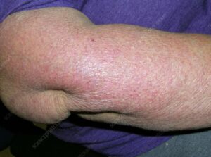
- Gottron’s Papules: Raised, pink, scaly patches that track directly down the fingers and/or populate the elbows and knees [cite: 2033, 2055, 2111].
- Gottron’s Sign: A flat, violaceous (purplish) discoloration stretching across the knuckles, elbows, or knees [cite: 2034, 2112].
- The V-Neck and “Shawl” Layout: Sun-sensitive (photodistributed) red-to-violet rashes form distinct geometric patterns across your upper body, including the “shawl sign” wrapping the upper back and shoulders, and a matching rash across the upper chest [cite: 2028, 2056, 2120].
- The Hand Architecture Clues: Look closely at your hands and fingers. DM targets the capillary beds, causing prominent telangiectasias (spider veins) along the nail folds, jagged or ragged cuticles, and a rough, lateral thickening of the finger skin known as “mechanic’s hands” [cite: 2030, 2036, 2058].
- The Myopathy Boundary: When muscle inflammation is present, it patterns itself symmetrically, selectively weakening the proximal muscle groups closest to the center of your body—specifically the triceps in the arms and the quadriceps in the thighs [cite: 2048, 2114, 2119]. This makes simple tasks like combing your hair or rising from a chair exceptionally difficult [cite: 2049].
Solutions I Can Try at Home
Because dermatomyositis is driven by an internal, immune-mediated process, home support focuses on blocking environmental triggers and safely maintaining your physical joint mobility [cite: 2060, 2091, 2092].
- Implement Strict Daily UV Protection: Ultraviolet light directly accelerates the immune signaling that fuels your skin rashes [cite: 2061]. Apply a broad-spectrum sunscreen daily, wear tightly woven long sleeves, and protect your neck and chest [cite: 2092]. Avoid direct sun exposure during peak daylight hours.
- Engage in Structured Physical Therapy: Work closely with a physical therapist to execute gentle, regular movement routines [cite: 2091]. Maintaining consistent movement is crucial to prevent permanent joint tightening (contractures) and minimize muscle wasting (atrophy) during an active flare [cite: 2091].
- Never Vigorously Scrub Active Plaques: Avoid using rough washcloths or harsh exfoliating scrubs on your knuckles or your scalp. Because the inflammation sits beneath the surface layer, aggressive scrubbing breaks your skin barrier lines, intensifies localized itching, and worsens tissue stress [cite: 2027, 2035, 2087].
When Should I See a Dermatology Provider?
Dermatomyositis is a complex condition that can easily mask itself as contact dermatitis, systemic lupus, or severe psoriasis [cite: 2074, 2075, 2076]. Seeking professional clinical triage early ensures you get a precise diagnosis through an office skin biopsy to check your tissue layers under a microscope and rule out lookalikes [cite: 2065].
Seek Professional Help if You Notice These Warning Signs:
- Sudden Eyelid Discoloration or Joint Rashes: You develop a new, purplish flush across your eyelids (heliotrope rash) or scaly pink patches tracking down your knuckles, requiring an immediate diagnostic evaluation [cite: 2032, 2033, 2113].
- New Symmetrical Muscle Weakness: You experience unprompted, matching weakness in your shoulders or thighs, find it difficult to lift your arms above your head, or struggle to stand up from a sitting position [cite: 2048, 2049].
- The Emergence of Lung or Swallowing Signals: Your skin or muscle changes are accompanied by internal warning flags, such as unexplained shortness of breath, a persistent dry cough, or sudden difficulty swallowing food [cite: 2046, 2050].
- Painful Palmar Papules and Fingertip Ulcers: You notice painful bumps on your palms or small open sores breaking out on your fingertips, which can indicate a highly severe variant known as MDA5 dermatomyositis that carries a risk of rapidly progressive interstitial lung disease and requires immediate emergency triage [cite: 2052, 2053].
Frequently Asked Questions
- Q: What extensive laboratory and imaging workup is required to confirm DM and check my safety?
A: Establishing a baseline diagnosis requires a highly comprehensive, multi-system screening panel [cite: 2067]. Your provider will order targeted blood tests including an Antinuclear Antibody (ANA) screen, DM-specific anti-muscle antibodies (Anti-MDA-5, Anti Mi-2, and Anti SRP), and the anti-synthetase profile (Anti Jo-1) [cite: 2069, 2123, 2124, 2125, 2126]. This is paired with checking active muscle enzymes—specifically Creatinine and Aldolase—where elevated levels provide clear evidence of cellular muscle damage [cite: 2069, 2109, 2110]. To fully check your internal safety, your interprofessional team will execute baseline strength testing, an electromyogram (EMG), ultrasound or MRI of affected muscle groups, and checks for pulmonary and cardiac health [cite: 2066, 2067, 2070]. Adults will also undergo thorough cancer screenings [cite: 2068, 2093]. - Q: What is the primary oral prescription medication protocol and taper schedule?
A: To aggressively halt systemic inflammation and achieve rapid disease control, your provider will initiate systemic oral corticosteroid therapy (Prednisone), traditionally starting at a dose of 1 mg/kg/day [cite: 2088, 2096, 2127]. Once your muscle and skin symptoms are successfully stabilized, your medical team will execute a highly structured, very slow weaning protocol [cite: 2097]. Most patients can become completely muscle disease-free after 24 to 48 months of a careful, gradual steroid taper [cite: 2097]. Corticosteroid treatments are frequently paired with topical steroid or non-steroidal tacrolimus/pimecrolimus creams to provide localized skin and scalp relief [cite: 2092]. - Q: What advanced options exist if my skin disease is stubborn or resistant to steroids?
A: Because available skin therapies are frequently inadequate on their own—leaving many patients with persistent, itchy cutaneous plaques and stubborn scalp distress—your team may need to step up care [cite: 2087]. For severe, chronic, or resistant cases, your provider will introduce powerful systemic immunosuppressants and steroid-sparing agents to protect your body [cite: 2089]. These advanced interventions include oral antimalarials (hydroxychloroquine or chloroquine), methotrexate, cyclosporine, mycophenolate mofetil, azathioprine, JAK 1 inhibitors, TNF-α inhibitors, rituximab, or Intravenous Immunoglobulin (IVIG) therapy [cite: 2089, 2090, 2128].
The long-term outlook for the majority of dermatomyositis patients is highly favorable when treatment is initiated early, allowing for excellent muscle recovery and successful symptom control [cite: 2096]. Success relies on strict daily ultraviolet protection habits, complete adherence to your multi-year structured medication taper, finishing all required internal heart and lung safety checks, and maintaining a regular follow-up schedule every 4 to 6 months with your dedicated healthcare team to keep your body safe and healthy [cite: 2067, 2072, 2092, 2097].